The Recombinant Cleaved N-terminal GSDMD Monoclonal Antibody is a recombinant rabbit monoclonal antibody (clone C370274) against Cleaved N-terminal GSDMD. GSDMD is a member of the gasdermin family. Members of this family appear to play a role in regulation of epithelial proliferation. GSDMD has been suggested to act as a tumor suppressor. Produced by recombinant technology in Rabbit hosts and purified by Protein A affinity chromatography, this Rabbit IgG antibody is reactive against Human samples.
This antibody is suitable for WB, IF and ELISA applications. It has been validated by WB analysis of THP-1. The target is localized to the cytoplasm, cytosol, inflammasome, in response to a canonical inflammasome stimulus, such as nigericin, recruited to NLRP3 inflammasone with similar kinetics to that of uncleaved CASP1 precursor, cell membrane, multi-pass membrane protein, secreted, released in the extracellular milieu following pyroptosis, cytoplasm, cytosol.
Recombinant Human Cleaved N-terminal GSDMD protein
Applications:
WBIFELISA
Clonality:
Recombinant Monoclonal
Clone No:
C370274
Host:
Rabbit
Isotype:
Rabbit IgG
Target:
Cleaved N-terminal GSDMD
Conjugation:
Unconjugated
Observed MW:
35 kDa
Calculated MW:
53 kDa
Recommended Dilution:
WB 1:2000-10000; IF 1:200-1000; ELISA 1:5000-20000
UniProt:
P57764
Purification:
Protein A
Concentration:
0.2 mg/mL
Buffer:
PBS, 50% glycerol, 0.05% Proclin 300, 0.05% protein protectant.
Storage:
Store at -20°C valid for 12 months. Avoid freeze / thaw cycles.
Immunofluorescence analysis of un-treated (left) A549 and UV treated(right) A549 cell. 1,primary Antibody was 1:200(4℃ diluted at overnight). 2, Goat Anti Rabbit IgG (H&L) - Alexa Fluor 594 Secondary antibody was diluted at 1:1000(room temperature, 50min).3, Picture B: DAPI(blue) 10min.
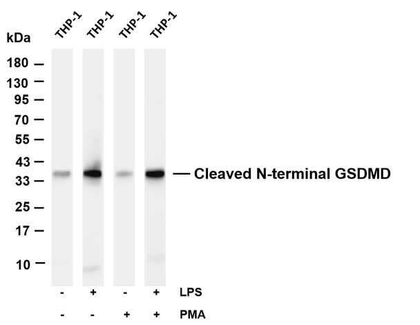
Various whole cell lysates were separated by 4-20% SDS- PAGE, and the membrane was blotted with anti-Cleaved N- terminal GSDMD antibody. The HRP-conjugated Goat anti- Rabbit IgG(H + L) antibody was used to detect the antibody. Lane 1: THP-1 Lane 2: THP-1 was treated with Lipopolysaccharides Lane 3: THP-1 was treated with Phorbol 12-myristate 13-acetate Lane 4: THP-1 was treated with Lipopolysaccharides and Phorbol 12-myristate 13-acetate Predicted band size: 53kDa Observed band size: 35kDa
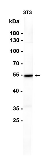
Various whole cell lysates were separated by 4-20% SDS- 4℃, PAGE, and the primary antibody was used at over night with a 1:5000 dilution. The Dylight 800-conjugated Goat anti-Rabbit antibody was used to detect the antibody. Lane1: HL60 Lane2: HUVEC Lane3: Human CAF Lane4: HCT116 Lane5: A549 Lane6: Jurkat Lane7: HT22 Lane8: THP-1 Lane9: HK-2 Lane10: PC-12 Lane11: 3T3- L1 Lane12: PC-3 Lane13: U-14 Lane14: 293F. Predicted band size: 35kDa Observed band size: 35kDa