Description
Aldehyde Dehydrogenase (ALDH) Fluorometric Activity Assay Kit
The Assay Genie Aldehyde Dehydrogenase (ALDH) Fluorometric Activity Assay Kit measures ALDH enzyme activity in serum, plasma, animal tissue homogenates, and cell lysates. ALDH converts substrate to NADH, which reacts with a fluorescent probe, and the signal is read at Ex/Em 535/587 nm. Researchers use the kit to quantify ALDH activity in liver and kidney function studies and across tissue and cell sample types.
Aldehyde dehydrogenase (ALDH) is the enzyme that oxidizes aldehydes to their corresponding acids. In the main pathway of alcohol metabolism, alcohol dehydrogenase (ADH) oxidizes alcohol to acetaldehyde, and NADH-dependent ALDH then oxidizes acetaldehyde to acetic acid. Because ALDH acts downstream of ADH, its activity determines how efficiently acetaldehyde is cleared from the system.
Researchers measure ALDH activity to study alcohol metabolism and to address liver and renal function, the research area listed for this kit. The kit is intended for research use only and is not for clinical diagnosis, a restriction stated in the manual. Documented sample types include serum, plasma, animal tissue, and cell lysates, including rat liver, mouse kidney, mouse liver, mouse lung, mouse brain, mouse spleen, mouse plasma, and Jurkat cells.
The assay is a fluorometric enzymatic method. ALDH acting on the substrate converts NAD+ to NADH, and NADH then reacts with a fluorescent probe under the action of enzymes to form a fluorescent product. Standard, sample, and control wells are each mixed with reaction working solution or buffer, then chromogenic working solution is added. After mixing and a 5 min reaction at room temperature with shading light, fluorescence is read at an excitation wavelength of 535 nm and an emission wavelength of 587 nm.
Serum and plasma are assayed directly. If they are not tested on the same day, they can be stored at -80°C for one month. Tissue and cell samples are washed in cold PBS (0.01 M, pH 7.4), homogenized in normal saline (0.9% NaCl) at 4°C, and centrifuged at 10000×g for 10 min to remove insoluble material. The supernatant is kept on ice, and its protein concentration is determined separately before the activity calculation.
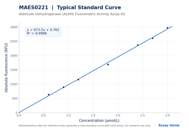
The kit reports an analytical sensitivity of 0.06 µmol/L and a detection range of 0.06–3 µmol/L. Intra-assay CV is 4.0% on the product page, and the manual's replicate data range from 3.8% to 4.2%. Inter-assay CV is 8.4% on the product page, and the manual's data range from 7.9% to 8.8%. Recovery at 0.8, 1.5, and 2.5 µmol/L was 99% to 101%, with an average of 100%.
| Product Name: | Aldehyde Dehydrogenase (ALDH) Fluorometric Activity Assay Kit |
| SKU: | MAES0221 |
| Synonyms: | ALDH |
| Assay Type: | Enzyme activity |
| Detection Method: | Fluorometric |
| Detection Instrument: | Fluorescence microplate reader (Ex/Em = 535 nm/587 nm) |
| Sample Types: | Serum, plasma, animal tissue, cells |
| Assay Time: | 20 min |
| Research Area: | Liver and renal function |
| Intended Use: | Research use only |
| Sensitivity: | 0.06 µmol/L |
| Detection Range: | 0.06–3 µmol/L |
| Intra-assay CV: | 4.0% |
| Inter-assay CV: | 8.4% |
| Average Recovery: | 100% (individual recoveries 99–101%) |
- Fluorometric detection, read at Ex/Em 535 nm/587 nm on a fluorescence microplate reader
- Detection range 0.06–3 µmol/L with a sensitivity of 0.06 µmol/L
- Assay time of 20 min
- Validated for serum, plasma, animal tissue, and cell samples
- Intra-assay CV 4.0% and inter-assay CV 8.4%
- Includes a standard curve preparation scheme (0–3 µmol/L) and calculation method
ALDH acts on the kit substrate and converts NAD+ to NADH. Under the action of the enzyme reagent, NADH reacts with a fluorescent probe to form a fluorescent product. ALDH activity is calculated from the fluorescence intensity measured at Ex/Em 535 nm/587 nm, using the sample well, the control well, and a standard curve.
- Measuring ALDH activity in rodent tissue homogenates, including liver, kidney, lung, brain, and spleen
- Assessing ALDH activity in mouse and rat liver tissue for liver function studies
- Quantifying ALDH activity in plasma or serum samples
- Measuring ALDH activity in Jurkat cell lysates
- Comparing ALDH activity across tissue types or experimental conditions
| Component | Quantity | Storage |
| Reagent 1: Buffer Solution | 20 mL (1 vial) | -20°C, 12 months |
| Reagent 2: Coenzyme Powder | 2 vials | -20°C, 12 months, protect from light |
| Reagent 3: Substrate | 0.3 mL (1 vial) | -20°C, 12 months, protect from light |
| Reagent 4: Chromogenic Agent | 0.2 mL (1 vial) | -20°C, 12 months, protect from light |
| Reagent 5: Enzyme Reagent Powder | 2 vials | -20°C, 12 months, protect from light |
| Reagent 6: Standard Powder | 2 vials | -20°C, 12 months, protect from light |
| Black microplate | 96 wells | No storage requirement |
| Plate sealer | 2 pieces | No storage requirement |
| Sample layout sheet | 1 piece | No storage requirement |
- Fluorescence microplate reader (Ex/Em = 535 nm/587 nm)
- Micropipettor
- Centrifuge capable of 10000 × g
- Double-distilled water
- Normal saline (0.9% NaCl)
- PBS (0.01 M, pH 7.4)
- Homogenizer (Dounce homogenizer for tissue; ultrasonic cell disruptor for cells)
- Ice
- Protein concentration assay for normalizing tissue and cell lysates
Serum/plasma
- Serum and plasma: detect directly.
- If not detected on the same day, store serum or plasma at -80°C for up to one month.
- Dilution guidance (reference only): mouse plasma, dilution factor 3–5.
- Diluent: normal saline (0.9% NaCl) or PBS (0.01 M, pH 7.4). For other sample types, run a pretest to confirm the dilution factor.
Tissue
- Harvest the amount of tissue needed for each assay (initial recommendation 20 mg).
- Wash tissue in cold PBS (0.01 M, pH 7.4).
- Homogenize 20 mg tissue in 180 µL normal saline (0.9% NaCl) with a Dounce homogenizer at 4°C.
- Centrifuge at 10000×g for 10 min to remove insoluble material. Collect the supernatant and keep it on ice for detection.
- Determine the protein concentration of the supernatant with a separate protein assay.
- Dilution guidance (reference only, 10% homogenate): rat liver 150–160; mouse kidney 90–150; mouse liver 150–180; mouse lung 60–90; mouse brain 30–40; mouse spleen 60–150.
- Diluent: normal saline (0.9% NaCl) or PBS (0.01 M, pH 7.4). For other sample types, run a pretest to confirm the dilution factor.
Cells (adherent or suspension)
- Harvest the number of cells needed for each assay (initial recommendation 1×10^6 cells).
- Wash cells with PBS (0.01 M, pH 7.4).
- Homogenize 1×10^6 cells in 200 µL normal saline (0.9% NaCl) with an ultrasonic cell disruptor at 4°C.
- Centrifuge at 10000×g for 10 min to remove insoluble material. Collect the supernatant and keep it on ice for detection.
- Determine the protein concentration of the supernatant with a separate protein assay.
- Dilution guidance (reference only): Jurkat cell, dilution factor 8–12.
- Diluent: normal saline (0.9% NaCl) or PBS (0.01 M, pH 7.4). For other sample types, run a pretest to confirm the dilution factor.
Reagent preparation
- Equilibrate all reagents to room temperature before use.
- Coenzyme working solution: dissolve one coenzyme vial in 1 mL double-distilled water and mix well. Store at 2–8°C for 2 days, protected from light.
- Reaction working solution (prepare fresh, use within 6 hours): for 1928 µL, mix 1770 µL buffer solution, 150 µL coenzyme working solution, and 8 µL substrate.
- Enzyme working solution: dissolve one enzyme reagent vial in 100 µL double-distilled water and keep on ice. Store at 2–8°C for 1 day, protected from light.
- Chromogenic working solution (prepare fresh, use within 6 hours): for 301 µL, mix 280 µL double-distilled water, 16 µL chromogenic agent, and 5 µL enzyme working solution.
- 300 µmol/L standard: dissolve one standard vial in 1.6 mL double-distilled water and mix well. Store at 4°C for 2 days, protected from light.
- 3 µmol/L standard: dilute 10 µL of 300 µmol/L standard with 990 µL double-distilled water. Prepare fresh.
Standard curve preparation
- Dilute the 3 µmol/L standard with double-distilled water as follows (standard volume / water volume): 0 µmol/L: 0 µL / 200 µL; 0.6 µmol/L: 40 µL / 160 µL; 0.9 µmol/L: 60 µL / 140 µL; 1.2 µmol/L: 80 µL / 120 µL; 1.8 µmol/L: 120 µL / 80 µL; 2.4 µmol/L: 160 µL / 40 µL; 2.7 µmol/L: 180 µL / 20 µL; 3 µmol/L: 200 µL / 0 µL.
- Prepare a fresh set of standards for each assay and discard working dilutions after use.
Sample preparation
- Serum and plasma: assay directly. If not assayed the same day, store at -80°C for up to 1 month.
- Tissue: wash tissue in cold PBS (0.01 M, pH 7.4). Homogenize 20 mg tissue in 180 µL normal saline (0.9% NaCl) with a Dounce homogenizer at 4°C. Centrifuge at 10000 × g for 10 min. Keep the supernatant on ice.
- Cells: wash cells with PBS (0.01 M, pH 7.4). Homogenize 1 × 10^6 cells in 200 µL normal saline (0.9% NaCl) with an ultrasonic cell disruptor at 4°C. Centrifuge at 10000 × g for 10 min. Keep the supernatant on ice.
- Determine protein concentration of tissue and cell supernatants for the calculation.
Tissue and cell samples: ALDH activity (U/g prot) = (F Sample − F Control − b) ÷ a ÷ T ÷ C_pr × f Serum (plasma) samples: ALDH activity (U/L) = (F Sample − F Control) ÷ a ÷ T × f
- F Sample: fluorescence intensity of the sample well.
- F Control: fluorescence intensity of the control well.
- b: intercept of the standard curve (y = ax + b), where y is the absolute fluorescence value of each standard (mean blank subtracted) and x is concentration in µmol/L.
- a: slope of the standard curve.
- T: incubation time of the reaction, 5 min.
- C_pr: protein concentration of the tissue or cell sample, g prot/L.
- f: dilution factor of the sample before the test.
- Units: for tissue and cells, 1 unit is the amount of ALDH in 1 g of tissue or cell protein that produces 1 µmol NADH per minute at 37°C. For serum or plasma, 1 unit is the amount of ALDH in 1 L of liquid sample that produces 1 µmol NADH per minute at 37°C.
Which sample types can be tested with this kit?
The kit measures ALDH activity in serum, plasma, animal tissue, and cell samples. The manual provides dilution guidance for rat liver, mouse kidney, mouse liver, mouse lung, mouse brain, mouse spleen, mouse plasma, and Jurkat cells. Other sample types require a pretest to confirm the dilution factor.
How should the reagents and prepared solutions be stored?
Kit components are stored at -20°C for 12 months, protected from light. Coenzyme working solution and enzyme working solution are stored at 2–8°C for 2 days and 1 day, respectively, protected from light. Reaction working solution and chromogenic working solution must be prepared on the spot and used within 6 hours.
What should I do if my sample is outside the detection range?
The detection range is 0.06–3 µmol/L. If the concentration is not within this range, perform an extra dilution or concentration of the sample. Refer to the dilution guidance for tissue and cell samples as a starting point.
What are the key points for avoiding assay problems?
Avoid bubbles when adding reaction working solution. Do not mix reagents from different kits. For small reagent volumes, centrifuge before use so that sufficient amounts are obtained.
What instrument settings and assay time are required?
Read fluorescence on a fluorescence microplate reader at an excitation wavelength of 535 nm and an emission wavelength of 587 nm, using a black microplate. After adding chromogenic working solution, mix for 3 s and react at room temperature with shading light for 5 min before reading. The total assay time is 20 min.
| Storage: | -20°C, protected from light |
| Shelf Life: | 12 months |
Notes
- For research use only; do not use in clinical diagnosis.
- Store all reagents strictly under the listed conditions and do not mix reagents from different kits.
- Centrifuge small-volume reagents before use so that sufficient amounts are obtained.
- Prepare the reaction working solution and chromogenic working solution on the spot and use within 6 hours.
- Avoid bubbles when adding the reaction working solution.