Description
Annexin V-AF488/7-AAD Apoptosis Kit
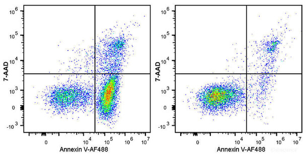
The Annexin V-AF488/7-AAD Apoptosis Kit detects apoptosis in suspension and adherent cells using Annexin V-AF488, which binds phosphatidylserine (PS) exposed on the outer leaflet of the apoptotic cell membrane. 7-AAD is included as a nuclear dye that enters late apoptotic or necrotic cells with compromised membrane integrity. Used together, the two stains distinguish early apoptotic, late apoptotic/necrotic, and naked nuclei populations by flow cytometry or fluorescence microscopy.
Annexin V is a member of the annexin family that binds phosphatidylserine (PS) in a calcium-dependent manner. In viable cells PS is restricted to the inner leaflet of the plasma membrane; during apoptosis it is exposed on the outer leaflet, where Annexin V-conjugated fluorophores can bind.
Loss of membrane integrity in late apoptotic or necrotic cells allows 7-AAD to enter and stain DNA, while intact cells exclude it. Combining Annexin V with 7-AAD allows cells at different apoptotic stages to be distinguished. The manual validates the kit with Jurkat cells treated with 5 µM camptothecin for 4 h.
| Product name: | Annexin V-AF488/7-AAD Apoptosis Kit |
| SKU: | AKES051 |
| Application: | Annexin V apoptosis detection |
| Detection method: | Fluorometric (flow cytometry, fluorescence microscopy) |
| Sample type: | Suspension and adherent cells |
| Assay time: | 15–20 min staining incubation |
| Detection instrument: | Flow cytometer or fluorescence microscope |
| Dye type: | AF488 (Annexin V conjugate) and 7-AAD (nuclear dye) |
| Ex/Em: | 495/520 nm (AF488) |
| Channel set: | FITC (AF488); PerCP/Cy5.5 (7-AAD) |
| Other reagents required: | PBS, deionized water |
- Annexin V-AF488 binds PS in a calcium-dependent manner for early apoptosis detection
- 7-AAD stains DNA in cells with compromised membrane integrity
- Compatible with suspension and adherent cells
- Single-stain-compatible readout: AF488 in FITC channel, 7-AAD in PerCP/Cy5.5 channel
- Assay time of 15–20 min staining, with one-step or two-step protocol options
Annexin V-AF488 binds specifically to PS on the outer leaflet of apoptotic cell membranes and is detected in the FITC channel (Ex/Em 495/520 nm). 7-AAD is excluded by intact cells and enters late apoptotic or necrotic cells to stain DNA, detected in the PerCP/Cy5.5 channel. Annexin V-positive, 7-AAD-negative cells are early apoptotic; Annexin V and 7-AAD double-positive cells are necrotic or late apoptotic; and 7-AAD single-positive cells are naked nuclei.
- Quantification of early apoptotic, late apoptotic/necrotic, and necrotic cell populations by flow cytometry
- Dead cell discrimination during flow cytometric analysis
- Fluorescence microscopy detection of PS exposure on apoptotic cells
- Assessment of apoptosis induced by experimental treatments, such as the camptothecin-treated Jurkat cell model in the manual
| Component | Storage |
| Annexin V-AF488 Reagent | 2-8°C, protect from light |
| Annexin V Binding Buffer (10×) | 2-8°C |
| 7-AAD Reagent (100 µg/mL) | 2-8°C, protect from light |
- PBS
- Deionized water (for diluting 10× Annexin V Binding Buffer)
- Centrifuge capable of 300 ×g
- Vortex mixer
- Flow cytometer with FITC and PerCP/Cy5.5 channels, or fluorescence microscope
- Microcentrifuge or flow cytometry tubes
- Ice and a dark incubation setup
- Trypsin without EDTA for adherent cell harvest (recommended)
Reagent preparation
- Dilute Annexin V Binding Buffer (10×) with deionized water to 1× working solution before use.
One-step protocol
- Induce apoptosis with the reagent of interest. Collect cells, centrifuge at 300 ×g for 5 min, and discard the supernatant. Wash with PBS, gently resuspend, and count cells.
- Aliquot 1–5 × 10⁵ cells per tube. Centrifuge at 300 ×g for 5 min, discard the supernatant, wash with PBS, and discard the supernatant. Resuspend in 500 µL of 1× Annexin V Binding Buffer.
- Add 5 µL of Annexin V-AF488 and 5 µL of 7-AAD to each tube.
- Gently vortex and incubate at room temperature for 15–20 min in the dark.
- Analyze immediately. Otherwise, place cells on ice in the dark and analyze within 1 h.
Two-step protocol
- Induce apoptosis with the reagent of interest. Collect cells, centrifuge at 300 ×g for 5 min, and discard the supernatant. Wash with PBS, gently resuspend, and count cells.
- Aliquot 1–5 × 10⁵ cells per tube. Centrifuge at 300 ×g for 5 min, discard the supernatant, wash with PBS, and discard the supernatant. Resuspend in 100 µL of 1× Annexin V Binding Buffer.
- Add 2.5 µL of Annexin V-AF488 and 2.5 µL of 7-AAD to each tube. The manual notes that users should titrate reagents for specific cell models.
- Gently vortex and incubate at room temperature for 15–20 min in the dark.
- Add 400 µL of 1× Annexin V Binding Buffer to each tube and mix gently.
- Analyze immediately. Otherwise, place cells on ice in the dark and analyze within 1 h.