Description
Annexin V-APC/Cyanine7/DAPI Apoptosis Kit
The Annexin V-APC/Cyanine7/DAPI Apoptosis Kit stains apoptotic cells for flow cytometry or fluorescence microscopy. Annexin V-APC/Cyanine7 binds phosphatidylserine exposed on the outer leaflet of the apoptotic cell membrane, and DAPI marks cells with lost membrane integrity. Together they separate early apoptotic, late apoptotic or necrotic, and nuclei-only populations.
Annexin V is a member of the annexin family that binds phosphatidylserine (PS) in a calcium-dependent manner. The APC/Cyanine7-conjugated format, Annexin V-APC/Cyanine7, binds specifically to PS on the outer leaflet of the membrane of apoptotic cells. The kit also includes DAPI, a nuclear dye that binds DNA. Because DAPI enters cells only after membrane integrity is lost, the two reagents together distinguish cells at different apoptotic stages. The kit is designed for detecting apoptosis in both suspension and adherent cells.
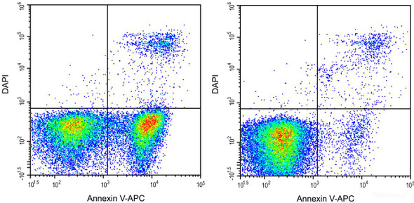
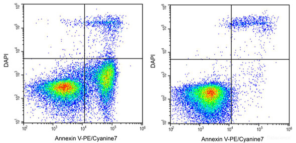
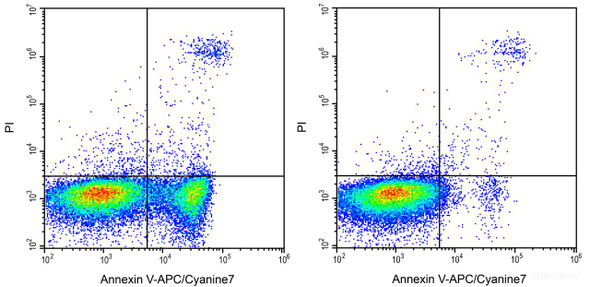
Researchers use Annexin V and DAPI co-staining to study apoptosis and cell death in cultures treated with reagents of interest. The manual illustrates this with Jurkat cells treated with 5 µM camptothecin for 4 h. In that example, Annexin V-APC/Cyanine7 single-positive cells were classified as early apoptotic, Annexin V-APC/Cyanine7 and DAPI double-positive cells as necrotic or late apoptotic, and DAPI single-positive events as naked nuclei. Separating these populations allows investigators to distinguish early apoptosis from late apoptosis and necrosis within the same sample.
Staining is performed on washed cells resuspended in 1× Annexin V Binding Buffer, prepared by diluting the 10× buffer with deionized water. Annexin V-APC/Cyanine7 and DAPI are added to each tube, and the cells are gently vortexed and incubated at room temperature for 15 to 20 min in the dark. Annexin V-APC/Cyanine7 is detected in the APC/Cy7 channel, with listed excitation/emission of 650; 760/780. The manual prefers the Pacific Blue channel for DAPI detection. Flow cytometry is the listed detection method, and fluorescence microscopy is also supported.
Suitable samples are cell suspensions and adherent cell cultures. Cells are washed with PBS and counted, and 1 to 5 × 10^5 cells are used per tube. Mechanical damage during adherent cell harvest should be minimized. Trypsin without EDTA is preferred because EDTA affects Annexin V binding to PS; if EDTA-containing trypsin is used, cells must be washed thoroughly. Suspension cells generated after apoptosis induction should be combined with the adherent cells. Stained samples should be analyzed immediately, or kept on ice in the dark and analyzed within 1 h, and extended exposure to direct light should be avoided.
| Product Name: | Annexin V-APC/Cyanine7/DAPI Apoptosis Kit |
| SKU: | AKES063 |
| Application: | Annexin V detection of apoptosis |
| Detection Method: | Fluorometric; flow cytometry or fluorescence microscopy |
| Sample Type: | Cell samples (suspension and adherent cells) |
| Assay Time: | 40 min |
| Detection Instrument: | Flow cytometer |
| Dye Type: | APC/Cyanine7 (Annexin V); DAPI (nuclear dye) |
| Ex/Em: | 650; 760/780 |
| Channel Set: | APC/Cy7 (Annexin V); Pacific Blue preferred for DAPI |
| Other Reagents Required: | PBS, deionized water |
- Dual-readout apoptosis staining: Annexin V-APC/Cyanine7 (phosphatidylserine) plus DAPI (membrane integrity)
- Detection by flow cytometry or fluorescence microscopy
- Suitable for suspension and adherent cell samples
- One-step and two-step staining protocols
- Approximately 40 min assay time with about 15 to 20 min room temperature incubation
- Stored at 2-8°C, protected from light, for 12 months
Annexin V-APC/Cyanine7 binds specifically to PS on the outer leaflet of apoptotic cell membranes and is detected in the APC/Cy7 channel by flow cytometry or fluorescence microscopy. DAPI enters cells that have lost membrane integrity and stains DNA. Annexin V-positive, DAPI-negative cells are early apoptotic; cells positive for both Annexin V and DAPI are necrotic or late apoptotic; DAPI-only single-positive events are naked nuclei.
- Quantifying early apoptosis, late apoptosis or necrosis, and naked nuclei in cell populations by flow cytometry
- Imaging apoptotic cells by fluorescence microscopy
- Evaluating apoptosis induced by experimental treatments in suspension or adherent cultures, for example Jurkat cells treated with 5 µmol/L camptothecin for 4 h
- Discriminating dead cells during flow cytometric analysis
| Component | Storage |
| Annexin V-APC/Cyanine7 Reagent | 2-8°C, protect from light |
| Annexin V Binding Buffer (10×) | 2-8°C |
| DAPI Reagent (25 µg/mL) | 2-8°C or -20°C, protect from light |
- PBS
- Deionized water (for diluting Annexin V Binding Buffer)
- Flow cytometer with APC/Cy7 and Pacific Blue channels, or fluorescence microscope
- Centrifuge capable of 300 ×g
- Vortex mixer
- Ice and light-protected storage for samples
- Trypsin without EDTA for adherent cell harvest (see cautions)
Suspension cells
- Induce apoptosis with the reagents of interest. Collect the cell culture, centrifuge at 300 × g for 5 min, and discard the supernatant.
- Add PBS to wash the cells, resuspend gently, and count the cells.
- Split the cell suspension into tubes at 1 to 5 × 10^5 cells per tube. Centrifuge at 300 × g for 5 min, discard the supernatant, add PBS to wash, and discard the supernatant again.
- One-step process: resuspend the cells in 500 µL of 1× Annexin V Binding Buffer. Two-step process: resuspend the cells in 100 µL of 1× Annexin V Binding Buffer.
- Prepare 1× Annexin V Binding Buffer by diluting Annexin V Binding Buffer (10×) with deionized water before use.
- One-step process: add 5 µL of Annexin V-APC/Cyanine7 and 5 µL of DAPI to each tube. Two-step process: add 2.5 µL of Annexin V-APC/Cyanine7 and 2.5 µL of DAPI to each tube. Titrating the reagents for specific cell models is recommended.
- Gently vortex the cells and incubate at room temperature for 15 to 20 min in the dark.
- Two-step process only: add 400 µL of 1× Annexin V Binding Buffer to each tube and mix gently.
- Analyze the cells immediately with appropriate instrument settings. Otherwise, place the cells on ice in the dark and analyze within 1 h.
Adherent cells
- Collect the suspension cells generated after apoptosis induction and detect them together with the subsequently collected adherent cells.
- Detach adherent cells while avoiding mechanical damage as much as possible.
- Use trypsin without EDTA where possible. If trypsin containing EDTA is used, wash the cells thoroughly after harvesting to remove EDTA.
- Continue with the suspension cell procedure from the centrifugation and washing steps, using the one-step or two-step staining process as required.
Reagent preparation
- Dilute Annexin V Binding Buffer (10×) with deionized water to 1×Annexin V Binding Buffer before use.
One-step process
- Induce apoptosis in cells with the reagent of interest. For suspension cells, collect cultures, centrifuge at 300 ×g for 5 min, and discard the supernatant. Wash with PBS, resuspend gently, and count cells.
- Split the cell suspension into tubes at 1–5×10⁵ cells per tube. Centrifuge at 300 ×g for 5 min, discard the supernatant, wash with PBS, and discard the supernatant again.
- Resuspend cells in 500 µL of 1×Annexin V Binding Buffer.
- Add 5 µL of Annexin V-APC/Cyanine7 and 5 µL of DAPI to each tube.
- Vortex gently and incubate at room temperature for 15–20 min in the dark.
- Analyze immediately. Otherwise, place cells on ice in the dark and analyze within 1 h.
Two-step process
- Induce apoptosis and prepare cells as in the one-step process, washing with PBS.
- Split cells at 1–5×10⁵ cells per tube, centrifuge at 300 ×g for 5 min, discard the supernatant, and wash with PBS.
- Resuspend cells in 100 µL of 1×Annexin V Binding Buffer.
- Add 2.5 µL of Annexin V-APC/Cyanine7 and 2.5 µL of DAPI to each tube.
- Vortex gently and incubate at room temperature for 15–20 min in the dark.
- Add 400 µL of 1×Annexin V Binding Buffer to each tube and mix gently.
- Analyze immediately. Otherwise, place cells on ice in the dark and analyze within 1 h.
Which sample types are compatible with this kit?
The kit is developed to detect apoptosis in suspension cells and adherent cells. Adherent cells should be harvested with care, and suspension cells collected after induction should be analyzed together with them.
How should the kit and reagents be stored?
The kit should be stored at 2 to 8°C, protected from light, and used within 12 months. The manual advises avoiding freeze/thaw cycles.
What should I do if the signal is outside the expected range?
The manual does not define an expected signal range. It states that control settings are critical to experimental results, and it recommends titrating the reagents for optimal performance in specific models.
What can interfere with Annexin V staining?
EDTA in trypsin affects Annexin V binding to phosphatidylserine, so EDTA-free trypsin is preferred. Extended exposure to direct light can quench the fluorophores, and samples should be analyzed promptly after staining.
Which instrument settings should be used?
Annexin V-APC/Cyanine7 is detected in the APC/Cy7 channel, with listed Ex/Em of 650; 760/780. DAPI is detected in the Pacific Blue channel, which the manual prefers over the DAPI channel.
How long does the assay take?
The listed assay time is 40 min. The staining incubation is 15 to 20 min at room temperature in the dark.
| Storage: | 2-8°C, protect from light |
| Expiration: | 12 months |
| Shipping: | Ice bag |
| Handling: | Avoid freeze/thaw cycles |
Cautions
- For Research Use Only. Not for use in diagnostic or therapeutic procedures.
- When detecting adherent cells, collect the suspension cells produced after apoptosis induction and analyze them together with the adherent cells.
- Minimize mechanical damage during adherent cell digestion. Avoid EDTA-containing trypsin, because EDTA interferes with Annexin V binding to phosphatidylserine. If EDTA-containing trypsin is used, wash cells thoroughly after harvest.
- Analyze samples as soon as possible after staining to avoid increases in apoptosis or necrosis.
- Avoid extended exposure of samples to direct light to prevent fluorophore quenching.
- Wear a lab coat and disposable gloves during experiments.