Description
Annexin V-PE/Cyanine5.5 Reagent
Annexin V-PE/Cyanine5.5 Reagent (AKES017) is a PE/Cyanine5.5-conjugated Annexin V probe for identifying apoptotic cells. Annexin V binds phosphatidylserine (PS) exposed on the outer leaflet of the apoptotic cell membrane in a calcium-dependent manner. Used with a membrane-impermeant DNA dye such as PI or DAPI, it separates early apoptotic, necrotic or late apoptotic, and nuclei-only populations by flow cytometry or fluorescence microscopy. For research use only.
Annexin V is a member of the annexin protein family. It binds phosphatidylserine (PS) in a calcium-dependent manner, and it binds PS specifically on the outer leaflet of the apoptotic cell membrane. This reagent uses Annexin V conjugated to PE/Cyanine5.5, so the bound protein carries a fluorescent label that can be read directly on the cell surface. Because the signal reports PS exposure at the membrane, it marks cells that have entered apoptosis rather than measuring the full apoptotic process. The reagent is for research use only.
Researchers use Annexin V-based staining to identify apoptotic cells in cultures exposed to experimental treatments. The kit is designed to separate cells at different apoptotic stages by pairing Annexin V with a membrane-impermeant DNA dye, either propidium iodide (PI) or DAPI. In the supplied example, Jurkat cells treated with 5 µM camptothecin for 4 h showed Annexin V single-positive cells, which were early apoptotic, and Annexin V and DAPI double-positive cells, which were necrotic or late apoptotic. DAPI single-positive events were naked nuclei.
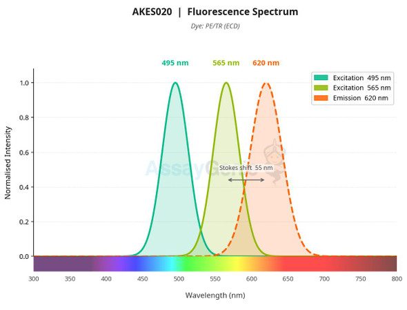
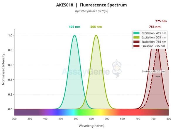
The assay is a fluorometric, cell-based stain. Cells are resuspended in Annexin V Binding Buffer, then incubated with 5 µL of Annexin V-PE/Cyanine5.5 Reagent and 5 µL of either PI or DAPI per tube. After gentle vortexing, the mixture is incubated for 15 to 20 min at room temperature in the dark. Samples are read by flow cytometry or fluorescence microscopy. The listed excitation and emission values are 495, 565, and 675/690, and the recommended channel set on the flow cytometer is PerCP/Cy5.5.
The protocol is written for suspension cells, and it also applies to adherent cells after harvest. Cells should be centrifuged at 300 × g for 5 min and washed with PBS before staining. For adherent cultures, suspension cells generated during apoptosis induction must be collected and analyzed together with the adherent cells. Mechanical damage from digestion should be minimized, and trypsin solutions should avoid EDTA because EDTA interferes with Annexin V binding to PS. Stained samples should be analyzed promptly and protected from direct light to limit fluorophore quenching.
| Product Name: | Annexin V-PE/Cyanine5.5 Reagent |
| SKU: | AKES017 |
| Pack Size: | 50 Tests, 200 Tests |
| Application: | Annexin V detection of apoptotic cells |
| Detection Method: | Fluorometric; flow cytometry or fluorescence microscopy |
| Sample Type: | Cell samples (suspension and adherent cells) |
| Assay Time: | 40 min |
| Detection Instrument: | Flow cytometer; fluorescence microscope |
| Dye Type: | PE/Cyanine5.5 |
| Channel Set: | PerCP/Cy5.5 |
| Other Reagents Required: | Annexin V Binding Buffer, PBS, deionized water, DNA dye (PI or DAPI) |
- Annexin V conjugated to PE/Cyanine5.5 for detection of PS exposure on apoptotic cells
- Compatible with flow cytometry (PerCP/Cy5.5 channel set) and fluorescence microscopy
- Assay time of 40 min; staining incubation of 15 to 20 min at room temperature in the dark
- Multiplexing with PI or DAPI to distinguish early apoptotic, necrotic/late apoptotic, and naked nuclei populations
- Calcium-dependent binding requires 1× Annexin V Binding Buffer during staining
- Storage at 2 to 8°C, protected from light, with a 12-month shelf life
Annexin V-PE/Cyanine5.5 binds specifically to PS on the outer leaflet of apoptotic cell membranes in a calcium-dependent manner. Bound conjugate is detected by flow cytometry or fluorescence microscopy using the PE/Cyanine5.5 fluorophore. Co-staining with PI or DAPI, which is excluded from intact membranes, separates early apoptotic from necrotic or late apoptotic cells.
- Identification of early apoptotic cells by PS exposure in suspension or harvested adherent cell populations
- Flow cytometric quantification of apoptotic subpopulations when combined with PI or DAPI
- Fluorescence microscopy detection of apoptotic cells
- Validation of apoptosis induction in cell-based experiments, for example Jurkat cells treated with 5 µM camptothecin for 4 h
| Component | Quantity | Storage |
| Annexin V-PE/Cyanine5.5 Reagent | 50 Tests: 125 µL × 2; 200 Tests: 500 µL × 2 | 2 to 8°C, protect from light |
- Flow cytometer or fluorescence microscope with appropriate filter sets
- Centrifuge capable of 300 × g
- 1× Annexin V Binding Buffer (prepared from 10× concentrate; 500 µL per sample)
- PBS for washing
- Deionized water
- DNA dye: PI or DAPI (5 µL per tube)
- Vortex mixer and ice bath
- Microcentrifuge tubes or flow cytometry tubes
- Apoptosis-inducing reagents of interest
Cells (suspension)
- Induce apoptosis in suspension cells with the reagents of interest.
- Collect the cell culture, centrifuge at 300 × g for 5 min, and discard the supernatant.
- Add PBS to wash the cells, resuspend gently, and count the cells.
- Split the cell suspension into tubes, using 1–5 × 10^5 cells per tube. Centrifuge at 300 × g for 5 min and discard the supernatant.
- Add PBS to wash the cells, then discard the supernatant.
- Add 500 µL of 1 × Annexin V Binding Buffer to resuspend the cells.
- Add 5 µL of Annexin V-PE/Cyanine5.5 Reagent and 5 µL of DNA dye (PI or DAPI) to each tube.
- Gently vortex the cells and incubate at room temperature for 15–20 min in the dark.
- Analyze immediately with appropriate instrument settings. Otherwise, place the cells on ice in the dark and analyze within 1 h.
Cells (adherent)
- Induce apoptosis with the reagents of interest.
- Collect the suspension cells generated after induction and combine them with the subsequently harvested adherent cells for analysis.
- Minimize mechanical damage during digestion of adherent cells.
- Avoid trypsin digestion solution containing EDTA where possible, because EDTA affects Annexin V binding to phosphatidylserine.
- If trypsin containing EDTA is used, wash the cells thoroughly after harvesting to remove EDTA.
- Wash, count, and resuspend the cells as described for suspension cells, then stain with Annexin V-PE/Cyanine5.5 Reagent and the DNA dye in 1 × Annexin V Binding Buffer.
- Incubate for 15–20 min at room temperature in the dark, then analyze immediately or on ice in the dark within 1 h.
Staining procedure
- Induce apoptosis in cells with the reagent of interest. For suspension cells, collect cultures, centrifuge at 300 × g for 5 min, discard the supernatant, wash with PBS, resuspend gently, and count. For adherent cells, collect detached suspension cells and adherent cells together, and minimize mechanical damage during digestion.
- Aliquot 1 to 5 × 10^5 cells per tube. Centrifuge at 300 × g for 5 min, discard the supernatant, wash with PBS, centrifuge again, and discard the supernatant.
- Resuspend the cell pellet in 500 µL of 1× Annexin V Binding Buffer.
- Add 5 µL of Annexin V-PE/Cyanine5.5 Reagent and 5 µL of DNA dye (PI or DAPI) to each tube.
- Gently vortex and incubate at room temperature for 15 to 20 min in the dark.
- Analyze immediately with appropriate instrument settings. If analysis is delayed, place cells on ice in the dark and analyze within 1 h.
What sample types can be used with this reagent?
The reagent is validated for cells, including suspension cells and adherent cells harvested after induction of apoptosis. For adherent cultures, detached suspension cells should be collected and analyzed together with the harvested adherent cells.
How should the reagent be stored?
The product information lists storage at 2–8°C for 12 months, protected from light. The manual specifies storage at 2–8°C in the dark for one year and advises avoiding freeze/thaw cycles.
Does EDTA interfere with the assay?
Yes. EDTA affects the binding of Annexin V to phosphatidylserine, so trypsin solutions without EDTA are preferred. If EDTA-containing trypsin is used, cells should be washed thoroughly after harvesting to remove it.
Which instrument settings apply?
Flow cytometry is the listed detection instrument, with a PerCP/Cy5.5 channel set and listed Ex/Em values of 495, 565, and 675/690. Fluorescence microscopy can also be used. Use proper machine settings for analysis.
How long does the assay take, and when should samples be read?
Incubation is 15–20 min at room temperature in the dark. The product information lists an assay time of 40 min. Analyze stained cells immediately, or place them on ice in the dark and analyze within 1 h.
| Storage Temperature: | 2 to 8°C |
| Storage Conditions: | Protect from light (shading light); avoid freeze/thaw cycles |
| Shelf Life: | 12 months |
| Shipping: | Ice bag |
Cautions
- For research use only. Not for diagnostic or therapeutic use.
- Avoid EDTA in trypsin or other digestion solutions where possible, as EDTA interferes with Annexin V binding to PS. If EDTA-containing trypsin is used, wash cells thoroughly after harvest.
- Minimize mechanical damage when harvesting adherent cells.
- Analyze cells as soon as possible after staining to avoid an increase in apoptotic and necrotic cells.
- Avoid extended exposure of stained samples to direct light to protect fluorophores from quenching.
- Wear a lab coat and disposable gloves during the procedure.