The Anti-AMPK alpha 1/2 Monoclonal Antibody (CAB27740) is an unconjugated rabbit monoclonal antibody developed for reliable detection of the catalytic alpha 1 and alpha 2 subunits of AMP-activated protein kinase (AMPK), the master cellular energy sensor that is activated when the AMP/ATP ratio rises. AMPK regulates key metabolic enzymes and protects cells from ATP depletion by switching off ATP-consuming biosynthetic pathways, making it a central target in metabolism, diabetes, obesity and cancer research. This antibody is supplied as a kit with Bradford Reagent for optional total protein quantification and accurate sample normalization.
This antibody is validated for use in WB, IHC-P, IF/ICC, ELISA applications and has demonstrated reactivity against Human, Mouse, Rat samples.
AMPK alpha 1 and alpha 2 (encoded by PRKAA1 and PRKAA2) belong to the ser/thr protein kinase family and are the catalytic subunits of the 5'-AMP-activated protein kinase (AMPK). AMPK is a cellular energy sensor conserved in all eukaryotic cells. The kinase activity of AMPK is activated by stimuli that increase the cellular AMP/ATP ratio. AMPK regulates the activities of a number of key metabolic enzymes through phosphorylation and protects cells from stresses that cause ATP depletion by switching off ATP-consuming biosynthetic pathways. Alternatively spliced transcript variants encoding distinct isoforms have been observed.
Purification Method
Affinity purification
Gene ID
5562/5563
UniProt
Q13131/P54646
Buffer Information
Store at -20°C. Avoid freeze / thaw cycles. Buffer: PBS with 0.09% sodium azide, 0.05% BSA, 50% glycerol, pH 7.3.
Bradford Reagent Storage
Store at room temperature for 1 year.
Protein Quantification (Optional)
To quantify total protein levels, use the Bradford Reagent included in this kit. Visit our Bradford Protein Assay Protocol to view the full protocol.
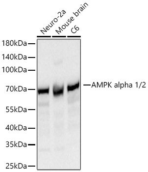
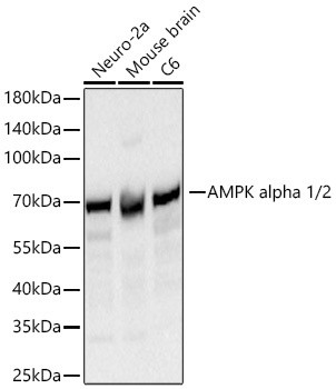
Western blot analysis of various lysates using Anti-AMPK alpha 1/2 Monoclonal Antibody (CAB27740) at 1:20000 dilution incubated overnight at 4°C. Secondary antibody: HRP-conjugated Goat anti-Rabbit IgG (H+L) at 1:10000 dilution. Lysates/proteins: 25 µg per lane. Blocking buffer: 3% nonfat dry milk in TBST. Detection: ECL Basic Kit. Exposure time: 30s.
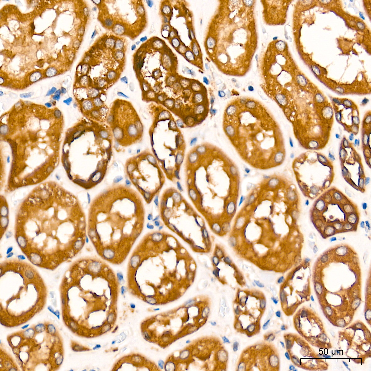
Immunohistochemistry analysis of paraffin-embedded Human kidney tissue using Anti-AMPK alpha 1/2 Monoclonal Antibody (CAB27740) at a dilution of 1:10000 (40x lens). High pressure antigen retrieval performed with 0.01 M Tris-EDTA buffer (pH 9.0) prior to IHC staining.
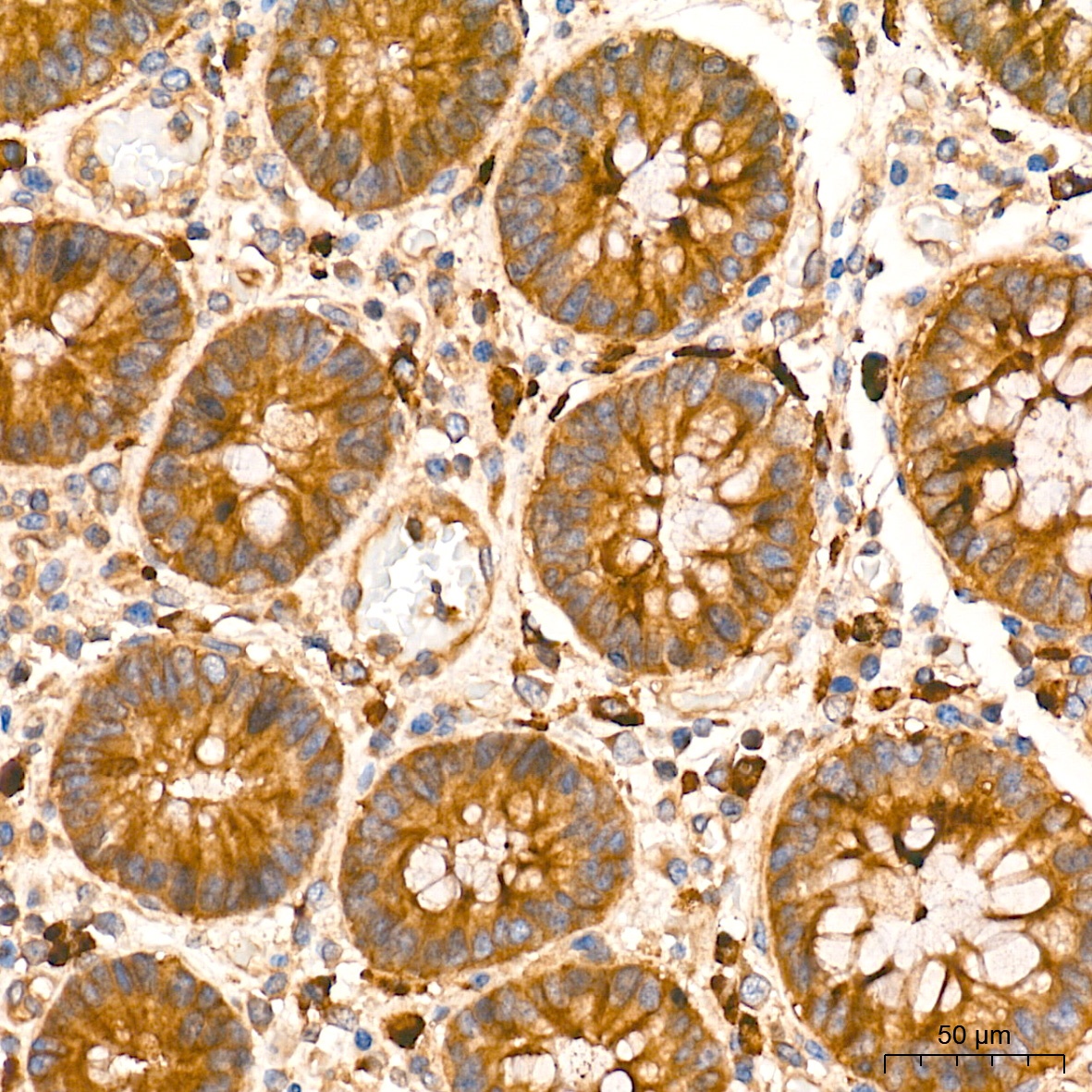
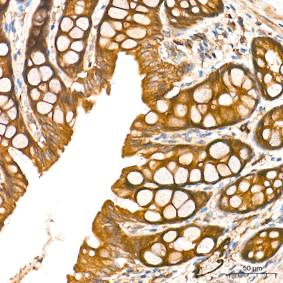
Immunohistochemistry analysis of paraffin-embedded Human colon tissue using Anti-AMPK alpha 1/2 Monoclonal Antibody (CAB27740) at a dilution of 1:10000 (40x lens). High pressure antigen retrieval performed with 0.01 M Tris-EDTA buffer (pH 9.0) prior to IHC staining.
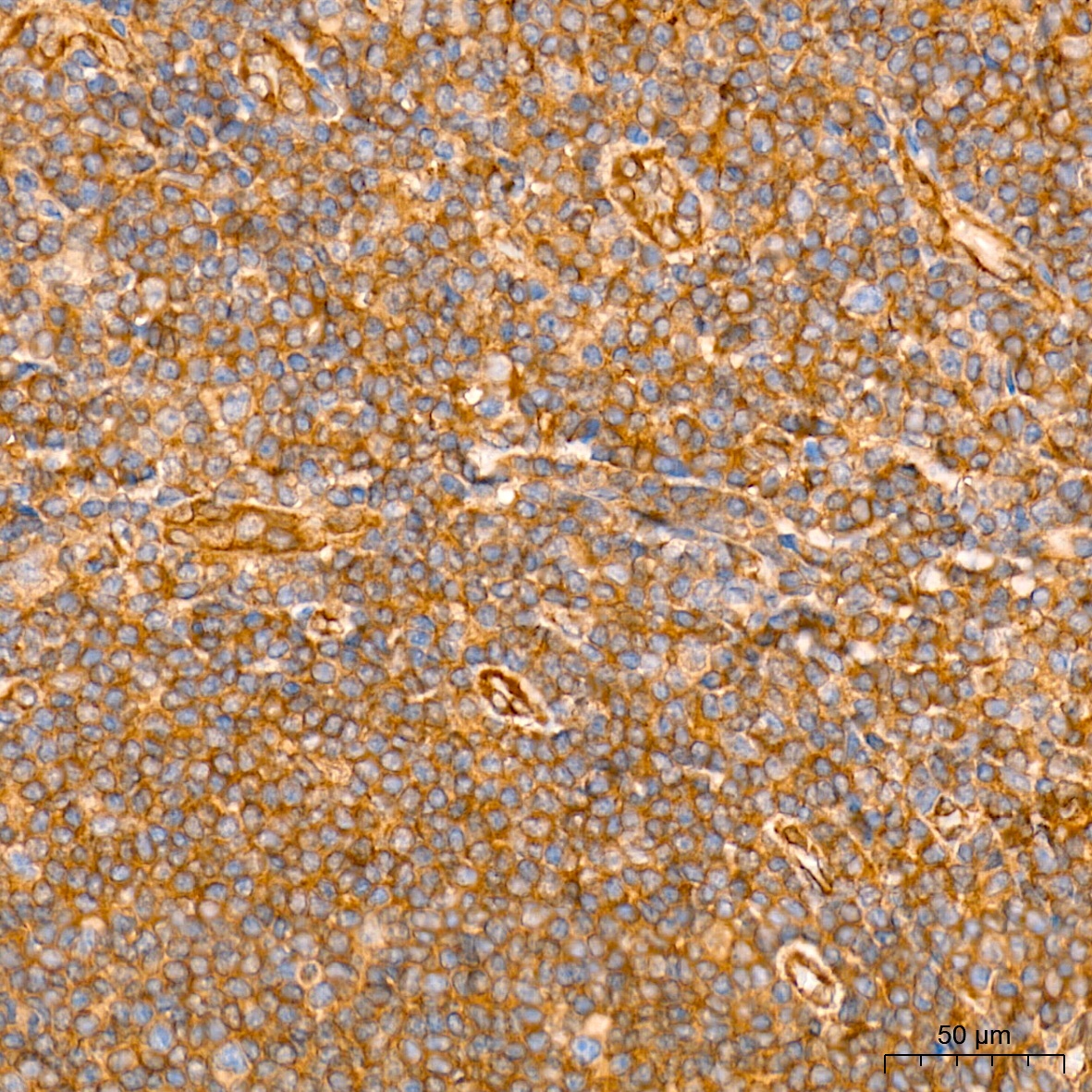
Immunohistochemistry analysis of paraffin-embedded Human tonsil tissue using Anti-AMPK alpha 1/2 Monoclonal Antibody (CAB27740) at a dilution of 1:10000 (40x lens). High pressure antigen retrieval performed with 0.01 M Tris-EDTA buffer (pH 9.0) prior to IHC staining.
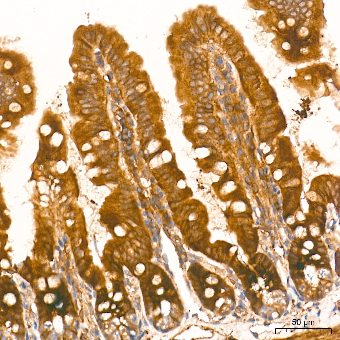
Immunohistochemistry analysis of paraffin-embedded Mouse colon tissue using Anti-AMPK alpha 1/2 Monoclonal Antibody (CAB27740) at a dilution of 1:10000 (40x lens). High pressure antigen retrieval performed with 0.01 M Tris-EDTA buffer (pH 9.0) prior to IHC staining.
Immunohistochemistry analysis of paraffin-embedded Rat colon tissue using Anti-AMPK alpha 1/2 Monoclonal Antibody (CAB27740) at a dilution of 1:10000 (40x lens). High pressure antigen retrieval performed with 0.01 M Tris-EDTA buffer (pH 9.0) prior to IHC staining.
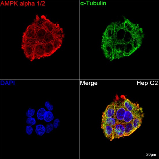
Confocal imaging of Hep G2 cells using Anti-AMPK alpha 1/2 Monoclonal Antibody (CAB27740, dilution 1:200) followed by a further incubation with Cy3 Goat Anti-Rabbit IgG (H+L) (dilution 1:500) (Red). The cells were counterstained with α-Tubulin Mouse mAb (dilution 1:400) followed by incubation with 488-conjugated Goat Anti-Mouse IgG (H+L) (dilution 1:500) (Green). DAPI was used for nuclear staining (Blue). Objective: 100x.
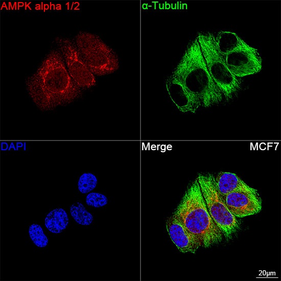
Confocal imaging of MCF7 cells using Anti-AMPK alpha 1/2 Monoclonal Antibody (CAB27740, dilution 1:200) followed by a further incubation with Cy3 Goat Anti-Rabbit IgG (H+L) (dilution 1:500) (Red). The cells were counterstained with α-Tubulin Mouse mAb (dilution 1:400) followed by incubation with 488-conjugated Goat Anti-Mouse IgG (H+L) (dilution 1:500) (Green). DAPI was used for nuclear staining (Blue). Objective: 100x.
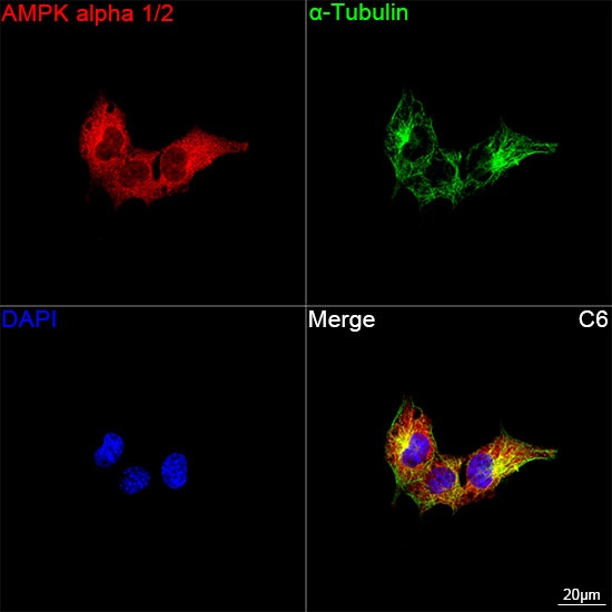
Confocal imaging of C6 cells using Anti-AMPK alpha 1/2 Monoclonal Antibody (CAB27740, dilution 1:200) followed by a further incubation with Cy3 Goat Anti-Rabbit IgG (H+L) (dilution 1:500) (Red). The cells were counterstained with α-Tubulin Mouse mAb (dilution 1:400) followed by incubation with 488-conjugated Goat Anti-Mouse IgG (H+L) (dilution 1:500) (Green). DAPI was used for nuclear staining (Blue). Objective: 100x.
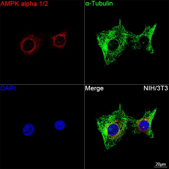
Confocal imaging of NIH/3T3 cells using Anti-AMPK alpha 1/2 Monoclonal Antibody (CAB27740, dilution 1:200) followed by a further incubation with Cy3 Goat Anti-Rabbit IgG (H+L) (dilution 1:500) (Red). The cells were counterstained with α-Tubulin Mouse mAb (dilution 1:400) followed by incubation with 488-conjugated Goat Anti-Mouse IgG (H+L) (dilution 1:500) (Green). DAPI was used for nuclear staining (Blue). Objective: 100x.