Description
Fluoride Resistant Acid Phosphatase (FRAP) Activity Assay Kit
The Assay Genie Fluoride Resistant Acid Phosphatase (FRAP) Activity Assay Kit (MAES0272) measures FRAP enzyme activity in serum, plasma, cells, and animal tissue samples. FRAP is not inhibited by fluoride ions, so activity can be measured in the presence of fluoride. The kit uses a colorimetric readout at 405 nm and is intended for Research Use Only.
Fluoride resistant acid phosphatase (FRAP) is the enzyme whose activity this kit measures. Its defining property is that it is not inhibited by fluoride ions, so its activity can be determined in the presence of fluoride. The kit includes a fluoride ion solution for this reason. FRAP catalyzes the conversion of its substrate to p-nitrophenol, and the amount of product formed reports the enzyme's activity in the sample.
The product page assigns this kit to the metabolic diseases research area. The listed sample types span animal, cell, and human-derived material: mouse liver, kidney, heart, and lung tissue homogenates, rat plasma, human and bovine serum, and HL-60 and 293T cells. This range supports comparative studies across tissues, species, and cell lines. The kit is for research use only and must not be used for clinical diagnosis.
The assay is a colorimetric, endpoint method. Standards or samples are combined with fluoride ion working solution and measuring working solution, which contains substrate working solution, then incubated at 37°C for 10 min. Chromogenic agent is added, and the optical density (OD) is read at 405 nm. The 405 nm peak corresponds to p-nitrophenol. A microplate reader with a 390-415 nm filter range is required, with 405 nm as the optimum wavelength. The total assay time is 25 min.
Serum, plasma, and tissue or cell samples are supported. Serum and plasma are tested directly, and tissue and cell samples are homogenized in 0.9% normal saline and clarified by centrifugation at 10000×g for 10 min. Supernatants are kept on ice until testing. Serum and plasma can be stored at -80°C for one month if they are not assayed on the day of collection. Sample dilution is required for many sample types, and the recommended dilution factors depend on the matrix.
Performance data come from the manual and the product page. The analytical sensitivity is 0.61 U/L, and the detection range is 0.61-49.37 U/L. Intra-assay CV is 3.6% on the product page, and the manual reports 3.5-3.8% across three human serum samples. Inter-assay CV is 8% on the product page, and the manual reports 7.6-8.2%. The average recovery is 102%, with individual recoveries of 101-104%.
| Product Name: | Fluoride Resistant Acid Phosphatase (FRAP) Activity Assay Kit |
| SKU: | MAES0272 |
| Synonyms: | FRAP |
| Sample Type: | Serum (plasma), cells, and animal tissue samples |
| Assay Type: | Enzyme activity |
| Detection Method: | Colorimetric |
| Detection Instrument: | Microplate reader (390–415 nm; optimum wavelength 405 nm) |
| Assay Time: | 25 min |
| Research Area: | Metabolic diseases |
| Sensitivity: | 0.61 U/L |
| Detection Range: | 0.61–49.37 U/L |
| Intra-assay CV: | 3.5%–3.8% (three human serum samples, 20 replicates each) |
| Inter-assay CV: | 7.6%–8.2% (three human serum samples, 20 runs in duplicate, three operators) |
| Mean Recovery: | 102% |
| Sample Volume per Well: | 20 µL |
| Detection Wavelength: | 405 nm (optimum); filter range 390–415 nm |
- Colorimetric readout: p-nitrophenol absorbance at 405 nm
- Fluoride ions do not inhibit FRAP activity
- Validated sample types: serum, plasma, cells, and animal tissue
- Assay time of 25 min on a microplate reader (390–415 nm; optimum 405 nm)
- Detection range 0.61–49.37 U/L; sensitivity 0.61 U/L
- Intra-assay CV 3.5%–3.8%; inter-assay CV 7.6%–8.2%
FRAP is not inhibited by fluoride ions. It catalyzes the hydrolysis of the substrate to produce p-nitrophenol, which has a maximum absorption peak at 405 nm. FRAP activity is calculated from the OD value measured at 405 nm against a p-nitrophenol standard curve. One unit (U) is defined as the amount of FRAP that produces 1 µmol p-nitrophenol per minute at 37°C.
- Measurement of FRAP activity in serum and plasma samples in metabolic disease research
- Quantification of FRAP activity in 10% mouse liver, kidney, heart, and lung tissue homogenates
- Measurement of FRAP activity in HL-60 and 293T cell lysates
- Comparative FRAP activity studies in assays that require fluoride-insensitive acid phosphatase activity
| Component | Quantity | Storage |
| Reagent 1: Buffer Solution | 20 mL x 1 vial | -20°C, 12 months |
| Reagent 2: Fluoride Ion Solution | 0.5 mL x 1 vial | -20°C, 12 months |
| Reagent 3: Substrate Solution | 1 mL x 1 vial | -20°C, 12 months, protect from light |
| Reagent 4: Chromogenic Agent | 14 mL x 1 vial | -20°C, 12 months |
| Reagent 5: 10 mmol/L Standard Solution | 1 mL x 1 vial | -20°C, 12 months, protect from light |
| Microplate | 96 wells x 1 | No requirement |
| Plate Sealer | 2 pieces | No requirement |
| Sample Layout Sheet | 1 piece | No requirement |
- Microplate reader (390–415 nm; optimum wavelength 405 nm)
- Incubator set to 37°C
- Double distilled water
- Normal saline (0.9% NaCl)
- PBS (0.01 M, pH 7.4) for sample washing
- Homogenizer (dounce homogenizer for tissue; ultrasonic cell disruptor for cells)
- Refrigerated centrifuge capable of 10000 × g
- Protein concentration assay for tissue and cell supernatants
- Ice and light-protective containers
Serum/plasma
- Detect serum or plasma directly without pretreatment.
- If samples are not assayed the same day, store serum or plasma at -80°C for up to one month.
- Serum and plasma are typically diluted before testing. Recommended dilution factors are 1-3 for human serum, 1-3 for bovine serum, and 5-7 for rat plasma.
- Use normal saline (0.9% NaCl) or PBS (0.01 M, pH 7.4) as the diluent. For other sample types, perform a pretest to confirm the dilution factor.
Tissue
- Harvest the amount of tissue needed for each assay. The initial recommendation is 20 mg.
- Wash the tissue in cold PBS (0.01 M, pH 7.4).
- Homogenize 20 mg of tissue in 180 µL of normal saline (0.9% NaCl) with a Dounce homogenizer at 4°C.
- Centrifuge at 10000×g for 10 min to remove insoluble material. Collect the supernatant and keep it on ice for detection.
- Determine the protein concentration of the supernatant separately, as required for the calculation.
- Dilute 10% mouse liver, kidney, heart, and lung tissue homogenates 3-7 times with normal saline (0.9% NaCl) before testing.
Cells
- Harvest the number of cells needed for each assay. The initial recommendation is 1×10^6 cells. This applies to adherent and suspension cells.
- Wash cells with PBS (0.01 M, pH 7.4).
- Homogenize 1×10^6 cells in 200 µL of normal saline (0.9% NaCl) with an ultrasonic cell disruptor at 4°C.
- Centrifuge at 10000×g for 10 min to remove insoluble material. Collect the supernatant and keep it on ice for detection.
- Determine the protein concentration of the supernatant separately, as required for the calculation.
- For HL-60 and 293T cells at 1×10^6 cells, the recommended dilution factor is 1.
Sample preparation
- Serum and plasma: assay directly. If not assayed the same day, store at -80°C for up to 1 month.
- Tissue: wash tissue in cold PBS, homogenize 20 mg tissue in 180 µL normal saline at 4°C, centrifuge at 10000 × g for 10 min, and keep the supernatant on ice.
- Cells: wash cells with PBS, homogenize 1 × 10⁶ cells in 200 µL normal saline at 4°C, centrifuge at 10000 × g for 10 min, and keep the supernatant on ice.
- Determine protein concentration of tissue and cell supernatants before calculation.
- Dilute samples as needed. Recommended dilution factors: 10% mouse liver, kidney, heart, or lung homogenate 3–7; rat plasma 5–7; human serum 1–3; bovine serum 1–3; 1 × 10⁶ HL-60 or 293T cells 1. Use normal saline or PBS as diluent. Pretest other sample types.
Reagent preparation
- Equilibrate all reagents to room temperature before use.
- Fluoride ion working solution: mix 5 µL Fluoride Ion Solution with 395 µL double distilled water. Prepare fresh and use within 1 hour.
- Substrate working solution: for each well, mix 4 µL Substrate Solution with 16 µL double distilled water. Prepare fresh and use within 1 hour.
- 0.5 mmol/L standard solution: dilute 45 µL of 10 mmol/L Standard Solution with 855 µL double distilled water. Keep on ice and protected from light. Use within 1 hour.
- Measuring working solution: for each well, mix 60 µL Buffer Solution with 20 µL substrate working solution. Prepare fresh.
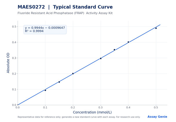
- Standard curve: dilute the 0.5 mmol/L standard with double distilled water to 0, 0.1, 0.15, 0.2, 0.3, 0.35, 0.4, and 0.5 mmol/L. Prepare fresh and discard after use.
Assay procedure
- Add 20 µL of each standard to standard wells, 20 µL of sample to sample wells, and 20 µL of sample to control wells.
- Add 20 µL fluoride ion working solution to each well.
- Add 80 µL measuring working solution to standard and sample wells. Add 80 µL Buffer Solution to control wells.
- Mix fully on the microplate reader and incubate at 37°C for 10 min.
- Add 100 µL Chromogenic Agent to each well.
- Mix fully and read OD at 405 nm.
Calculation
- Average duplicate readings for each standard. Subtract the mean blank OD (standard 0 mmol/L) from all standard readings to obtain absolute OD.
- Plot absolute OD (y) against standard concentration in mmol/L (x) and fit a linear standard curve (y = ax + b).
- Calculate serum or plasma FRAP activity (U/L) as (ΔA405 − b) ÷ a ÷ T × f × 1000, where ΔA405 = ODsample − ODcontrol, T = 10 min, and f = sample dilution factor.
- Calculate tissue or cell FRAP activity (U/gprot) as (ΔA405 − b) ÷ a ÷ Cpr ÷ T × f × 1000, where Cpr = protein concentration of the sample in gprot/L.
Serum (plasma) FRAP activity (U/L) = (ΔA405 -b)÷a÷T×f×1000 Tissue and cell FRAP activity (U/gprot) = (ΔA405 -b)÷a÷Cpr÷T×f×1000
- ΔA405 is the OD at 405 nm of the sample minus the OD of the control well. The control well contains sample and buffer solution in place of measuring working solution.
- a and b are the slope and intercept of the standard curve, y = ax + b, where y is the absolute OD value of each standard and x is its concentration in mmol/L. Absolute OD is the standard reading minus the mean OD of the blank (Standard 1).
- T is the reaction time, 10 min.
- Cpr is the protein concentration of the sample in gprot/L. It applies to tissue and cell samples only.
- f is the dilution factor of the sample before testing. Multiply by f to correct for any dilution applied during sample preparation.
- 1000 converts mmol/L to µmol/L (1 mmol/L = 1000 µmol/L).
- One unit is defined as the amount of FRAP that hydrolyzes the substrate to produce 1 µmol of p-nitrophenol in 1 min at 37°C. Results are reported in U/L for serum and plasma and in U/gprot for tissue and cell samples.
Which sample types can be tested with this kit?
The kit measures FRAP activity in serum, plasma, cells, and animal tissue samples. Serum and plasma are tested directly, while tissue and cell samples are homogenized in normal saline and clarified by centrifugation at 10000×g for 10 min.
How should samples and reagents be stored?
All reagents should be stored at -20°C for 12 months. Substrate Solution and Standard Solution should also be protected from light. Serum and plasma can be stored at -80°C for one month if they are not assayed the same day.
What should I do if my sample is outside the detection range?
The detection range is 0.61-49.37 U/L. If the concentration is not within this range, perform an extra dilution or concentration of the sample. For sample types not listed in the protocol, run a pretest to confirm the dilution factor.
What are the key points for accurate results?
Fluoride ions do not inhibit FRAP, so the fluoride ion working solution is part of the assay design. Prepare the fluoride ion working solution, substrate working solution, and 0.5 mmol/L standard solution fresh, and use them within 1 hour. Equilibrate all reagents to room temperature before use.
What instrument settings are required?
Use a microplate reader with a 390-415 nm wavelength range and read absorbance at 405 nm, the optimum wavelength. Incubate samples at 37°C for 10 min after mixing, then read the OD at 405 nm after adding chromogenic agent.
How long does the assay take?
The assay time is 25 min. This covers the 10 min incubation at 37°C and the subsequent addition of chromogenic agent and OD reading at 405 nm.
| Storage: | -20°C |
| Valid Period: | 12 months |
| Light Sensitivity: | Protect Substrate Solution and 10 mmol/L Standard Solution from light |
| Working Solutions: | Prepare fresh on the day of use; use within 1 hour |
Notes
- For Research Use Only. Not for use in clinical diagnosis or therapeutic procedures.
- Equilibrate all reagents to room temperature before use.
- Do not mix reagents from different kits.
- Centrifuge small reagent volumes briefly before use to ensure adequate recovery.
- Prepare fluoride ion working solution, substrate working solution, 0.5 mmol/L standard solution, and standard dilutions fresh and use within 1 hour.
- If a sample falls outside the detection range (0.61–49.37 U/L), dilute or concentrate the sample and repeat the assay.
- Run a pretest for sample types not listed in the dilution table.
- Wear a lab coat and gloves during use.