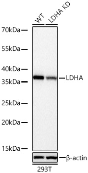
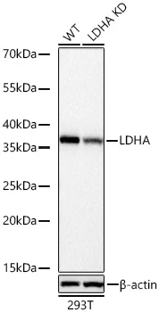
The LDHA Rabbit mAb (CAB28205) is an unconjugated rabbit monoclonal antibody (clone ARC77247) developed for reliable detection of lactate dehydrogenase A (LDHA), the enzyme that catalyzes the final step of anaerobic glycolysis. Its specificity has been confirmed by knockdown validation, with a clearly reduced LDHA signal in LDHA knockdown 293T cells compared with wild type cells. LDHA is widely studied in metabolism and cancer research, where elevated glycolytic activity (the Warburg effect) is a hallmark of many tumors. This antibody is supplied as a kit with Bradford Reagent for optional total protein quantification and accurate sample normalization.
This antibody is validated for use in WB, IF/ICC, ELISA applications and has demonstrated reactivity against Human, Mouse, Rat samples.
Recombinant protein (or fragment). This information is considered to be commercially sensitive.
Tested Applications:
WBIF/ICCELISA
Recommended Dilution:
WB
1:2000 - 1:15000
IF/ICC
1:200 - 1:400
ELISA
Recommended starting concentration is 1 µg/mL. Please optimize the concentration based on your specific assay requirements. For high-ratio antibody dilutions (≥1:10000), a sequential dilution method is strongly recommended to ensure measurement accuracy.
Synonyms:
LDHM, GSD11, PIG19, HEL-S-133P, LDHA
Positive Sample:
293T, U-87 MG, NIH/3T3, Rat liver
Cellular Localization:
Cytoplasm.
Calculated MW:
37kDa
Observed MW:
37kDa
The protein encoded by this gene catalyzes the conversion of L-lactate and NAD to pyruvate and NADH in the final step of anaerobic glycolysis. The protein is found predominantly in muscle tissue and belongs to the lactate dehydrogenase family. Mutations in this gene have been linked to exertional myoglobinuria. Multiple transcript variants encoding different isoforms have been found for this gene. The human genome contains several non-transcribed pseudogenes of this gene.
Purification Method
Affinity purification
Gene ID
3939
UniProt
P00338
Buffer Information
Store at -20°C. Avoid freeze / thaw cycles. Buffer: PBS with 0.09% sodium azide, 0.05% BSA, 50% glycerol, pH 7.3.
Bradford Reagent Storage
Store at room temperature for 1 year.
Protein Quantification (Optional)
To quantify total protein levels, use the Bradford Reagent included in this kit. Visit our Bradford Protein Assay Protocol to view the full protocol.
Western blot analysis of lysates from wild type (WT) and LDHA knockdown (KD) 293T cells using LDHA Rabbit mAb (CAB28205) at 1:5000 dilution incubated overnight at 4°C. Secondary antibody: HRP-conjugated Goat anti-Rabbit IgG (H+L) at 1:10000 dilution. Lysates/proteins: 25 µg per lane. Blocking buffer: 3% nonfat dry milk in TBST. Detection: ECL Basic Kit. Exposure time: 1s.
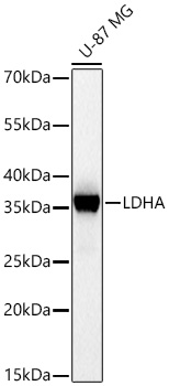
Western blot analysis of lysates from U-87 MG cells using LDHA Rabbit mAb (CAB28205) at 1:5000 dilution incubated overnight at 4°C. Secondary antibody: HRP-conjugated Goat anti-Rabbit IgG (H+L) at 1:10000 dilution. Lysates/proteins: 25 µg per lane. Blocking buffer: 3% nonfat dry milk in TBST. Detection: ECL Basic Kit. Exposure time: 1s.
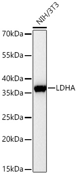
Western blot analysis of lysates from NIH/3T3 cells using LDHA Rabbit mAb (CAB28205) at 1:5000 dilution incubated overnight at 4°C. Secondary antibody: HRP-conjugated Goat anti-Rabbit IgG (H+L) at 1:10000 dilution. Lysates/proteins: 25 µg per lane. Blocking buffer: 3% nonfat dry milk in TBST. Detection: ECL Basic Kit. Exposure time: 30s.
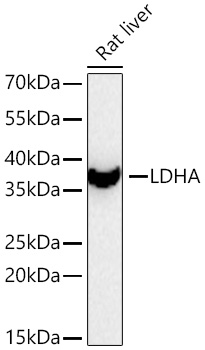
Western blot analysis of lysates from Rat liver using LDHA Rabbit mAb (CAB28205) at 1:5000 dilution incubated at room temperature for 1.5 hours. Secondary antibody: HRP-conjugated Goat anti-Rabbit IgG (H+L) at 1:10000 dilution. Lysates/proteins: 25 µg per lane. Blocking buffer: 3% nonfat dry milk in TBST. Detection: ECL Basic Kit. Exposure time: 30s.
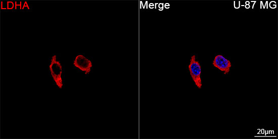
Confocal imaging of U-87 MG cells using LDHA Rabbit mAb (CAB28205, dilution 1:200) followed by a further incubation with Cy3-conjugated Goat anti-Rabbit IgG (H+L) (dilution 1:500) (Red). DAPI was used for nuclear staining (Blue). Objective: 100x.