Description
Leucine Aminopeptidase (LAP) Activity Assay Kit
The Assay Genie Leucine Aminopeptidase (LAP) Activity Assay Kit (MAES0190) measures LAP enzyme activity in serum, plasma, and animal tissue homogenates. LAP hydrolyzes L-leucine-4-nitroaniline to release p-nitroaniline, which is quantified by absorbance at 405 nm in a 96-well microplate format. The kit is used in research workflows that examine LAP activity in liver and hepatobiliary models.
Leucine aminopeptidase (LAP) is an enzyme that acts on the chromogenic substrate L-leucine-4-nitroaniline, hydrolyzing it to release p-nitroaniline. Measuring LAP activity therefore means measuring the rate at which this colored product accumulates. This Assay Genie kit quantifies LAP activity in animal tissue, serum, plasma, and other liquid samples. Activity is expressed in units, where one unit is the amount of enzyme that produces 1 µmol of p-nitroaniline per minute at 37 °C. The kit includes a substrate powder, an extracting solution, and a p-nitroaniline standard.
Researchers measure LAP activity in studies of liver and renal function, which is the research area listed for this kit. The kit has been applied to serum and plasma from human, dog, rat, and cynomolgus monkey, and to 10% tissue homogenates from rat spleen, liver, kidney, and lung, which supports comparative work across these species and organs. Serum and plasma are assayed directly, while tissue is washed in cold PBS and kept on ice during preparation. The kit is for research use only and is not intended for clinical diagnosis.
The assay is a colorimetric, two-read kinetic method performed in a 96-well microplate. The substrate working solution is prepared by dissolving the substrate powder in anhydrous ethanol, and it is kept on ice during use. Each sample well receives 10 µL of sample and 170 µL of extracting solution, then 20 µL of substrate working solution. The absorbance at 405 nm is read after a 5 s mix (A1), the plate is incubated at 37 °C for 10 min, and the absorbance is read again (A2). The change in optical density, ΔA = A2 − A1, reflects p-nitroaniline production. Standard wells do not change over time, so their A2 values are used to plot the standard curve.
Reported performance includes an analytical sensitivity of 5.2 U/L and a detection range of 5.2–201.8 U/L. Sensitivity was determined by adding two standard deviations to the mean OD of the zero standard, assayed 20 times, and converting that signal to concentration. Intra-assay CV ranged from 3.8% to 4.5% across three human serum samples, and inter-assay CV ranged from 6.0% to 7.0%. Recovery of p-nitroaniline at low, middle, and high concentrations averaged 99%, with individual recoveries of 97% to 101%.
| Product Name: | Leucine Aminopeptidase (LAP) Activity Assay Kit |
| SKU: | MAES0190 |
| Product Size: | 96 Assays |
| Sample Type: | Serum, plasma, animal tissue |
| Detection Method: | Colorimetric |
| Instrument: | Microplate reader (405 nm) |
| Assay Time: | 25 min |
| Research Area: | Enzymes; liver and renal function |
| Sensitivity: | 5.2 U/L |
| Detection Range: | 5.2-201.8 U/L |
| Intra-assay CV: | 4.1% |
| Inter-assay CV: | 6.4% |
| Sample Volume: | 10 µL |
| Wavelength: | 405 nm |
| Assay Time: | 25 min |
- Colorimetric detection of p-nitroaniline at 405 nm on a microplate reader
- Detection range 5.2-201.8 U/L with a stated analytical sensitivity of 5.2 U/L
- Serum and plasma tested directly; tissue homogenates prepared in extracting solution
- 10 µL sample volume per well
- Kinetic read: absorbance measured before and after 10 min at 37 °C
- Includes p-nitroaniline standard for a standard curve
LAP catalyzes the substrate L-leucine-4-nitroaniline to produce p-nitroaniline, which has a maximum absorption peak at 405 nm. LAP activity is calculated from the increase in OD value of the reaction system over 10 min at 37 °C, corrected for the sample blank reading taken before incubation.
- Quantification of LAP activity in animal serum and plasma
- Measurement of LAP activity in rodent liver, kidney, spleen, and lung tissue homogenates
- Comparative LAP activity studies across human, dog, rat, and cynomolgus monkey serum
- Hepatobiliary and liver injury research models
| Component | Quantity | Storage |
| Extracting Solution | 60 mL × 2 vials | 2-8 °C, 12 months |
| Substrate Powder | 2 vials | 2-8 °C, 12 months, protect from light |
| p-Nitroaniline Standard Powder | 1 vial | 2-8 °C, 12 months, protect from light |
| 96-well Microplate | 1 plate (96 wells) | No special storage requirement |
- Microplate reader capable of reading at 405 nm
- Micropipettor and tips
- Water bath or incubator set to 37 °C
- Centrifuge capable of 10000 × g
- Vortex mixer
- Ice
- Dounce-type tissue homogenizer (for tissue samples)
- Cold PBS, 0.01 M, pH 7.4 (for tissue washing)
- Double-distilled water
- Anhydrous ethanol (for substrate and standard stock preparation)
- Protein assay for tissue supernatant (user-supplied, required for U/g prot calculation)
Serum/plasma
- Detect serum or plasma directly without extraction.
- The recommended dilution factor is 1 for human, dog, rat, and cynomolgus monkey serum.
- The diluent is extracting solution.
- For sample types not listed, perform a pretest to confirm the dilution factor.
Tissue
- Harvest the amount of tissue needed for each assay (initial recommendation 20 mg).
- Wash the tissue in cold PBS (0.01 M, pH 7.4).
- Homogenize 20 mg of tissue in 180 µL of extracting solution with a Dounce homogenizer at 4 °C, yielding a 10% homogenate.
- Centrifuge at 10000 × g for 10 minutes to remove insoluble material.
- Collect the supernatant and keep it on ice for detection.
- Determine the protein concentration of the supernatant separately, as it is required for the tissue calculation.
- The recommended dilution factor is 1 for 10% rat spleen, liver, kidney, and lung homogenates.
- The diluent is extracting solution. For other tissue types, perform a pretest to confirm the dilution factor.
Reagent preparation
- Equilibrate all reagents to room temperature before use.
- Substrate working solution: dissolve one vial of substrate powder in 1.2 mL anhydrous ethanol and mix well. Keep on ice during use. Store at 2-8 °C for up to 7 days, protected from light.
- p-Nitroaniline 50 mmol/L stock: dissolve one vial of standard powder in 1 mL anhydrous ethanol and mix well.
- p-Nitroaniline 1 mmol/L standard solution: add 20 µL of 50 mmol/L stock to 980 µL extracting solution and mix. Prepare on the day of use, keep on ice, and store at 2-8 °C for up to 7 days protected from light.
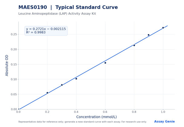
- Standard curve: prepare a fresh set of standards for each assay and discard working dilutions after use. Dilute the 1 mmol/L standard with extracting solution as follows: 0 (0 µL standard and 200 µL extracting solution), 0.2 mmol/L (40 µL and 160 µL), 0.3 mmol/L (60 µL and 140 µL), 0.4 mmol/L (80 µL and 120 µL), 0.6 mmol/L (120 µL and 80 µL), 0.8 mmol/L (160 µL and 40 µL), 0.9 mmol/L (180 µL and 20 µL), 1.0 mmol/L (200 µL and 0 µL).
Sample preparation
- Serum or plasma: assay directly.
- Tissue: wash tissue in cold PBS (0.01 M, pH 7.4). Homogenize 20 mg tissue in 180 µL extracting solution with a Dounce homogenizer at 4 °C. Centrifuge at 10000 × g for 10 min. Collect the supernatant and keep on ice.
- Determine the protein concentration of the tissue supernatant for normalization.
- Dilution: recommended dilution factor is 1 for human, dog, rat, and cynomolgus monkey serum, and for 10% rat spleen, liver, kidney, and lung homogenates. Use extracting solution as diluent. For other sample types, perform a pretest to confirm the dilution factor.
Assay procedure
- Standard wells: add 180 µL extracting solution, then 20 µL of each standard.
- Sample wells: add 10 µL sample and 170 µL extracting solution.
- Add 20 µL substrate working solution to sample wells.
- Mix for 5 s and read OD at 405 nm (A1).
- Incubate at 37 °C for 10 min, then read OD at 405 nm again (A2). Calculate ΔA = A2 − A1.
- Standard wells do not change in OD; plot the standard curve using the A2 reading of the standards.
- Avoid bubbles when transferring supernatant to the microplate.
Serum/plasma and other liquid samples: LAP activity (U/L) = (ΔA − b) ÷ a × V1 ÷ V2 ÷ T × 1000 × f Tissue samples: LAP activity (U/gprot) = (ΔA − b) ÷ a × V1 ÷ V2 ÷ T × 1000 × f ÷ Cpr
- ΔA = A2 − A1, the change in OD at 405 nm over the 10 min incubation.
- a and b are the slope and intercept of the standard curve y = ax + b, where y is the absolute OD and x is the p-nitroaniline concentration in mmol/L.
- V1 is the volume of substrate working solution added, 20 µL.
- V2 is the volume of sample added, 10 µL.
- T is the reaction time, 10 min.
- 1000 converts mmol to µmol (1 mmol = 1000 µmol).
- f is the dilution factor of the sample before testing. Multiply the result by f; a sample diluted before testing is corrected by this factor.
- Cpr is the protein concentration of the sample in g prot/L, used only for tissue samples.
- Results are reported in U/L for serum and plasma and in U/g prot for tissue.
What sample types can be used with this kit?
The kit measures LAP activity in animal tissue, serum, plasma, and other liquid samples. Serum and plasma are assayed directly, and tissue samples are homogenized and centrifuged before testing.
How should the kit components be stored?
Extracting solution, substrate powder, and p-nitroaniline standard powder are stored at 2–8 °C for 12 months, with the substrate and standard protected from light. The substrate working solution and 1 mmol/L p-nitroaniline standard solution are stored at 2–8 °C for 7 days, protected from light.
What should I do if my sample is outside the detection range?
The detection range is 5.2–201.8 U/L. If the concentration of a sample is not within this range, perform an additional dilution or concentration of the sample before testing.
Are there key steps to avoid assay errors?
Prevent bubbles from forming when the supernatant is transferred into the microplate. Results depend closely on reagents, operation, and environment, so follow the instructions strictly and run a pretest for any sample type not listed.
What instrument settings and assay time are required?
Read absorbance at 405 nm with a microplate reader. Add 10 µL of sample per well, and incubate the plate at 37 °C for 10 min between the two reads. The total assay time is 25 min.