Description
Mouse PHF8 ELISA Kit
Mouse PHF8 ELISA kits are used to detect PHD Finger Protein 8 (PHF8) in tissue homogenates and other biological samples. PHF8 is a histone lysine demethylase of the Jumonji C domain family that removes mono- and dimethyl groups from histone H3 lysine 9 and monomethyl groups from histone H4 lysine 20, marks associated with transcriptional repression. By removing these marks it acts as a transcriptional activator, and it plays a key role in cell cycle progression, being required for the G1-S transition, and in rDNA transcription by promoting RNA polymerase I activity. PHF8 is also required for brain development, partly by enhancing KDM5C expression with the transcription factor ARX. Variants in the human gene are associated with X-linked intellectual disability, and PHF8 is also studied in cancer.
| Product Name: | Mouse PHF8 (PHD Finger Protein 8) ELISA Kit |
| SKU: | AEKE02100 |
| Size: | 96 Assays |
| Reactivity: | Mouse |
| Synonyms: | ZNF422, MRXSSD, JHDM1F, Jumonji C Domain-Containing Histone Demethylase 1F, Histone lysine demethylase PHF8 |
| Assay Type: | Sandwich |
| Sensitivity: | 0.118 ng/mL |
| Range: | 0.32-20 ng/mL |
| Standard: | 20 ng/mL |
| Assay Length: | 3.5h |
| Sample Type: | Tissue Homogenates and Other Biological Fluids. |
| Kit Components: |
| ||||||||||||||||||||||||||||||||||||||||||||||||||||
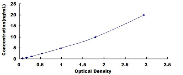
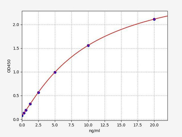
The test principle applied in this kit is Sandwich enzyme immunoassay. The microtiter plate provided in this kit has been pre-coated with an antibody specific to Mouse PHF8. Standards or samples are added to the appropriate microtiter plate wells then with a biotin-conjugated antibody specific to Mouse PHF8. Next, Avidin conjugated to Horseradish Peroxidase (HRP) is added to each microplate well and incubated. After TMB substrate solution is added, only those wells that contain Mouse PHF8, biotin-conjugated antibody and enzyme-conjugated Avidin will exhibit a change in color. The enzyme-substrate reaction is terminated by the addition of sulphuric acid solution and the color change is measured spectrophotometrically at a wavelength of 450nm ± 10nm. The concentration of Mouse PHF8 in the samples is then determined by comparing the OD of the samples to the standard curve.
| Standard Curve: |
| |||||||||||||||||||||||||||
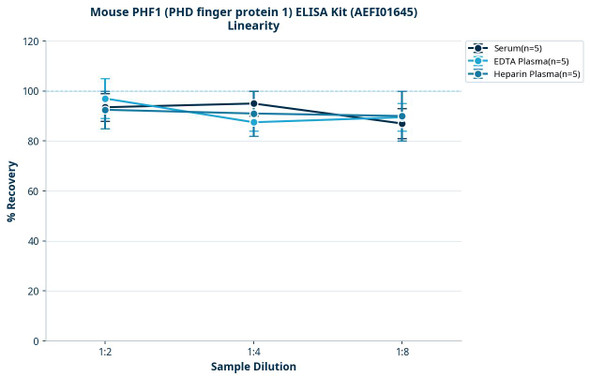
| Linearity: |
| |||||||||||||||||||||||||||
| Recovery: |
| |||||||||||||||||||||||||||
| Precision: | Intra-assay Precision (Precision within an assay):CV%<8% Three samples of known concentration were tested twenty times on one plate to assess intra-assay precision. Inter-assay Precision (Precision between assays):CV%<10% Three samples of known concentration were tested in forty separate assays to assess inter-assay precision. |
*Note:The below protocol is a sample protocol. Protocols are specific to each batch/lot. For the correct instructions please follow the protocol included in your kit.
| Step | Protocol |
| 1. | After the kit is equilibrated at room temperature, add 100 μL of Standard Working Buffer (gradually diluted according to the instructions) or 100 μL of sample to each well, and incubate at 37°C for 80 minutes. |
| 2. | Discard the liquid in the plate, add 200 μL 1× Wash Buffer to each well, and wash the plate 3 times. After pat it dry against clean absorbent paper, add 100 μL Biotinylated Antibody Working Solution (1×) to each well, incubate at 37°C for 50 minutes. |
| 3. | Discard the liquid in the plate, add 200 μL 1× Wash Buffer to each well, and wash the plate 3 times. After pat it dry against clean absorbent paper, add 100 μL 1× Streptavidin-HRP Working Solution to each well, incubate at 37°C for 50 minutes. |
| 4. | Discard the liquid in the plate, add 200 μL 1× Wash Buffer to each well, and wash the plate 5 times. After pat it dry against clean absorbent paper, add 90 μL TMB Substrate Solution to each well, incubate at 37°C for 20 minutes in the dark. |
| 5. | Add 50 μL Stop Solution to each well, shake plate on a plate shaker for 1 minute to mix. Record the OD at 450 nm immediately, calculation of the results. |
When carrying out an ELISA assay it is important to prepare your samples in order to achieve the best possible results. Below we have a list of procedures for the preparation of samples for different sample types.
| Sample Type | Protocol |
| Serum | Samples should be collected into a serum separator tube. After clotting for 2 hours at room temperature or overnight at 4°C, and then centrifuging at 1000 × g for 20 minutes. Assay freshly prepared serum immediately or store samples in aliquot at -20°C or -80°C for later use. Avoid repeated freeze-thaw cycles. |
| Plasma | Collect plasma using EDTA or heparin as an anticoagulant. Centrifuge samples at 1000 × g and 2-8°C for 15 minutes within 30 minutes of collection. Remove plasma and assay immediately or store samples in aliquot at -20°C or -80°C for later use. Avoid repeated freeze-thaw cycles. |
| Tissue homogenates | 1. Rinse the tissues in pre-cooled PBS to completely remove excess blood, and weigh them before homogenization. 2. Mince the tissues and homogenize in fresh lysis buffer (PBS for most tissues). Use a glass homogenizer on ice. 3. Ultrasound the suspension until the solution is clear. 4. Centrifuge for 5 minutes at 10000 × g, collect the supernatant and assay immediately or store at ≤ -20°C. |
| Cell lysates | 1. Wash adherent cells with PBS, detach with trypsin, and centrifuge at 1000 × g for 5 minutes. 2. Wash cells 3 times in PBS. 3. Resuspend cells in fresh lysis buffer at 10⁷ cells/mL. Ultrasound if necessary. 4. Centrifuge at 1500 × g for 10 minutes at 2-8°C to remove debris. Assay immediately or store at ≤ -20°C. |
| Urine | Collect mid-stream first urine of the day directly into a sterile container. Centrifuge to remove particulate matter. Assay immediately or aliquot and store at ≤ -20°C. Avoid repeated freeze-thaw cycles. |
| Saliva | Collect saliva using a collection device. Centrifuge at 1000 × g for 15 minutes at 2-8°C. Remove particulates and assay immediately or aliquot and store at ≤ -20°C. Avoid repeated freeze-thaw cycles. |
| Feces | Dry feces weighing more than 50 mg were collected. Wash with PBS (w:v = 1:9). Sonicate and centrifuge at 5000 × g for 10 minutes. Collect the supernatant and assay immediately. |
| CSF (Cerebrospinal fluid) | Remove particulates by centrifugation. Assay immediately or aliquot and store at ≤ -20°C. Avoid repeated freeze-thaw cycles. |
| Cell culture supernatant | Centrifuge samples at 1000 × g for 20 minutes. Collect the supernatant and assay immediately or store at -20°C or -80°C. Avoid repeated freeze-thaw cycles. |
| Uniprot ID: | Q80TJ7 |
| Research Area: | Signal Transduction |