The Recombinant CD13 Monoclonal Antibody is a recombinant rabbit monoclonal antibody (clone C111084) against CD13. Broad specificity aminopeptidase which plays a role in the final digestion of peptides generated from hydrolysis of proteins by gastric and pancreatic proteases. Also involved in the processing of various peptides including peptide hormones, such as angiotensin III and IV, neuropeptides, and chemokines. May also be involved the cleavage of peptides bound to major histocompatibility complex class II molecules of antigen presenting cells. Produced by recombinant technology in Rabbit hosts and purified by Protein A affinity chromatography, this Rabbit IgG antibody is reactive against Human, Mouse and Rat samples.
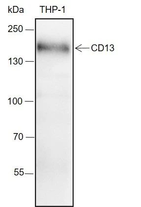
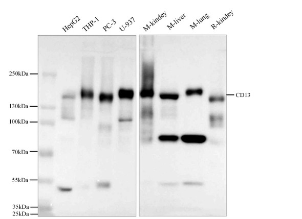
This antibody is suitable for WB, IHC, IF and FCM applications. It has been validated by WB analysis of THP-1, IHC analysis of Human breast, Human kidney, Mouse kidney, Rat kidney, IF analysis of THP-1, A375 and FCM analysis of THP-1. The target is localized to the cytoplasm, membrane.
Product Name:
Recombinant CD13 Monoclonal Antibody
SKU:
RMEA1085
Size:
50 µL, 100 µL
Antibody Type:
Recombinant Monoclonal Antibody
Synonyms:
Alanyl (membrane) aminopeptidase, Alanyl aminopeptidase, Aminopeptidase M, Aminopeptidase N, AMPN, ANPEP, AP M, AP N, AP-M, AP-N, APN, CD 13, CD13, CD13 antigen, CD13APN, GP150, hAPN, LAP, LAP 1, LAP1, Microsomal aminopeptidase, Myeloid plasma membrane glycoprotein CD13, P150, PEPN, PEPNhAPN
Reactivity:
Human, Mouse, Rat
Immunogen:
Recombinant human CD13 fragment
Applications:
WBIHCIFFCM
Clonality:
Recombinant Monoclonal
Clone No:
C111084
Host:
Rabbit
Isotype:
Rabbit IgG
Target:
CD13
Conjugation:
Unconjugated
Observed MW:
150 kDa
Calculated MW:
110 kDa
Recommended Dilution:
WB 1:500-2000; IHC 1:200-1000; IF 1:50-100; FCM 1:50-100
UniProt:
P15144
Purification:
Protein A
Concentration:
1 mg/mL
Buffer:
PBS, 50% glycerol, 0.05% Proclin 300, 0.05% protein protectant.
Storage:
Store at -20°C valid for 12 months. Avoid freeze / thaw cycles.
Flow cytometric analysis of human CD13 expression on THP-1 cells. Cells were stained with purified anti-Human CD13, then a Alexa Fluor 488-conjugated second step antibody. The histogram were derived from events with the forward and side light-scatter characteristics of intact cells.
Immunofluorescent analysis of (4% Paraformaldehyde) fixed THP-1 cells using anti-CD13 Monoclonal Antibody at dilution of 1:100.
Immunofluorescent analysis of (4% Paraformaldehyde) fixed A375 cells using anti-CD13 Monoclonal Antibody at dilution of 1:50.
Immunohistochemistry of paraffin-embedded Human breast using CD13 Monoclonal Antibody at dilution of 1:1000.
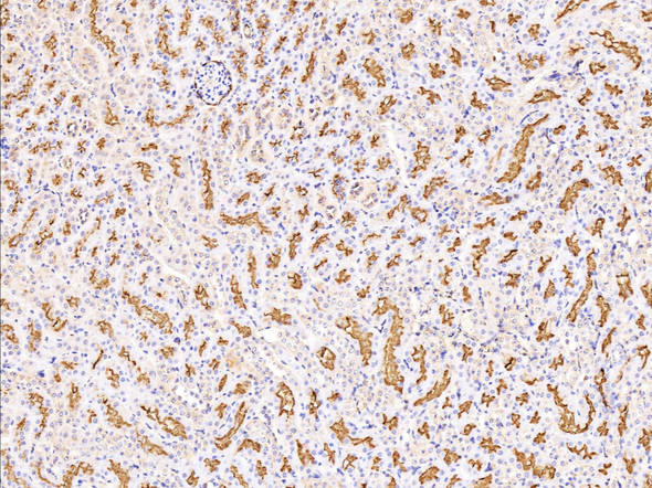
Immunohistochemistry of paraffin-embedded Human kidney using CD13 Monoclonal Antibody at dilution of 1:1000.
Immunohistochemistry of paraffin-embedded Mouse kidney using CD13 Monoclonal Antibody at dilution of 1:1000.
Immunohistochemistry of paraffin-embedded Rat kidney using CD13 Monoclonal Antibody at dilution of 1:1000.
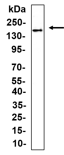
Western Blot with CD13 Monoclonal Antibody at dilution of 1:2000. Lane 1: THP-1 Observed-MW:150 kDa Calculated-MW:110 kDa