The Recombinant eIF3G Monoclonal Antibody is a recombinant rabbit monoclonal antibody (clone C111594) against eIF3G. eIF3 is a large translation initiation complex with 10 to 13 different subunits. eIF3A, eIF3B, eIF3C, eIF3E, eIF3F, and eIF3H are the core subunits critical for the function of this complex. eIF3 physically interacts with eIF4G, which may be responsible for the association of the 40S ribosomal subunit with mRNA. eIF3 also stabilizes the binding of Met-tRNAf.eIF2.GTP to the 40S ribosomal subunit and helps keep the integrity of the resulting complex upon addition of the 60S ribosomal subunit. Studies have shown that mTOR interacts with eIF3 directly. When cells are stimulated by hormones or mitogenic signals, mTOR binds to the eIF3 complex and phosphorylates S6K1. Produced by recombinant technology in Rabbit hosts and purified by Protein A affinity chromatography, this Rabbit IgG antibody is reactive against Human, Mouse and Rat samples.
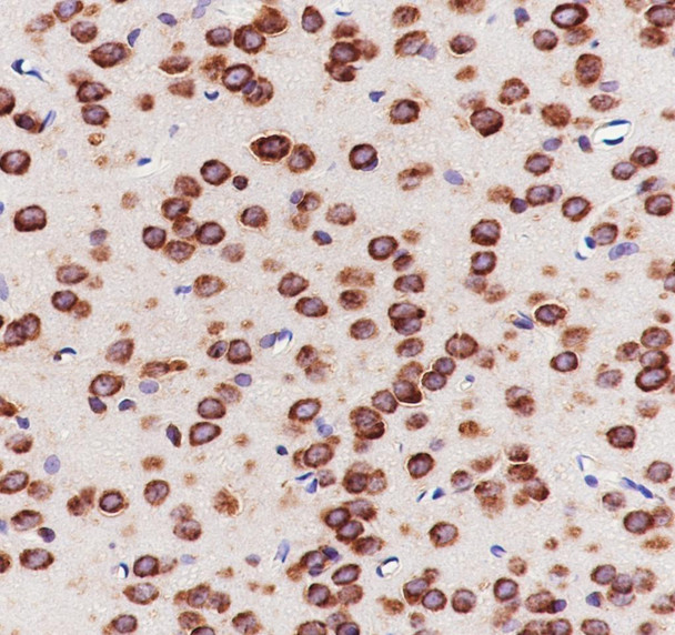
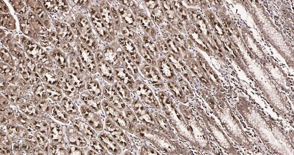
This antibody is suitable for WB, IHC, IF and FCM applications. It has been validated by WB analysis of K562, 293T, NIH-3T3, PC-12, IHC analysis of Human colon cancer, Mouse cerebrum, Rat cerebrum, IF analysis of 293T, K-562 and FCM analysis of K-562. The target is localized to the nucleus, cytoplasm.
Peptide. This information is proprietary to PTMab.
Applications:
WBIHCIFFCM
Clonality:
Recombinant Monoclonal
Clone No:
C111594
Host:
Rabbit
Isotype:
Rabbit IgG
Target:
eIF3G
Conjugation:
Unconjugated
Observed MW:
40 kDa
Calculated MW:
36 kDa
Recommended Dilution:
WB 1:1000-2000; IHC 1:50-100; IF 1:50-100; FCM 1:50
UniProt:
O75821
Purification:
Protein A
Concentration:
1 mg/mL
Buffer:
PBS, 50% glycerol, 0.05% Proclin 300, 0.05% protein protectant.
Storage:
Store at -20°C valid for 12 months. Avoid freeze / thaw cycles.
Flow cytometric analysis of human eIF3G expression on K-562 cells. Cells were stained with purified anti-Human eIF3G, then a Alexa Fluor 488-conjugated second step antibody. The histogram were derived from events with the forward and side light-scatter characteristics of intact cells.
Immunofluorescent analysis of (4% Paraformaldehyde) fixed 293T cells using anti-eIF3G Monoclonal Antibody at dilution of 1:100.
Immunofluorescent analysis of (4% Paraformaldehyde) fixed K-562 cells using anti-eIF3G Monoclonal Antibody at dilution of 1:100.
Immunohistochemistry of paraffin-embedded Human colon cancer using eIF3G Monoclonal Antibody at dilution of 1:100.
Immunohistochemistry of paraffin-embedded Mouse cerebrum using eIF3G Monoclonal Antibody at dilution of 1:100.
Immunohistochemistry of paraffin-embedded Rat cerebrum using eIF3G Monoclonal Antibody at dilution of 1:100.
Western Blot with eIF3G Monoclonal Antibody at dilution of 1:2000. Lane 1: K562, Lane 2: 293T, Lane 3: NIH-3T3, Lane 4: PC-12 Observed-MW:40 kDa Calculated-MW:36 kDa