Description
Acid Phosphatase (ACP) Activity Assay Kit
The Acid Phosphatase (ACP) Activity Assay Kit (MAES0024) measures ACP activity in serum, plasma, and tissue homogenates using a colorimetric p-nitrophenyl phosphate (PNPP) substrate. Acid phosphatase hydrolyzes PNPP to p-nitrophenol, which is read at 405 nm after alkaline stop. The kit is intended for Research Use Only and supports enzyme activity studies in liver, renal, and tissue research.
Acid phosphatase (ACP) is a phosphatase enzyme that catalyzes the hydrolysis of phosphate esters, releasing inorganic phosphate from phosphorylated substrates. Activity is measured as the rate at which the enzyme converts a chromogenic substrate into a detectable product, so the readout reflects enzyme function in the sample rather than total protein mass. For this kit, ACP activity is reported in units per liter (U/L) for serum and plasma and in units per gram of protein (U/g protein) for tissue homogenates. The kit is intended to measure ACP activity in serum, plasma, and tissue samples and is for research use only.
Researchers measure ACP activity in studies of liver and renal function, the research area listed for this kit. The supplied materials document rat liver, kidney, and spleen homogenates, along with plasma from mouse, rat, and human sources and human urine, as test matrices. Comparing ACP activity across these tissues and biofluids allows investigators to characterize enzyme levels in different organs under experimental conditions. A 10% Epipremnum aureum tissue homogenate is also listed, extending use to plant tissue studies.
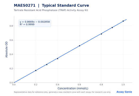
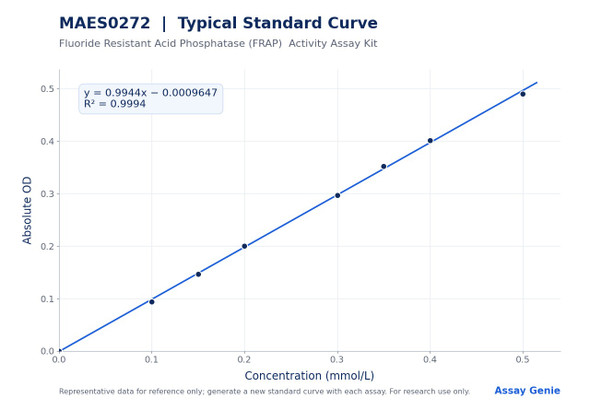
The assay uses disodium p-nitrophenyl phosphate (p-NPP), a widely used phosphatase chromogenic substrate. ACP converts p-NPP to p-nitrophenol, which is yellow under alkaline conditions and has a maximum absorption peak at 405 nm. Sample is combined with substrate working solution and incubated at 37°C for 10 min, after which chromogenic agent is added to each well. Absorbance is read at 405 nm on a microplate reader (400-415 nm), and a darker yellow product indicates higher ACP activity. A p-nitrophenol standard series from 0 to 0.5 mmol/L is run on each plate to generate the standard curve.
Serum and plasma are assayed directly. If they are not tested on the same day, they can be stored at -80°C for one month. Animal tissue is washed in cold PBS (0.01 M, pH 7.4), homogenized at 4°C, and centrifuged at 10000×g for 10 min at 4°C to remove insoluble material, and the supernatant is kept on ice until use. Samples require dilution before testing, with the factor depending on sample type. Dilution guidance is provided for listed matrices, and a pretest is recommended for other sample types. Substrate working solution should be used within one day, and substrate and standard should be protected from light.
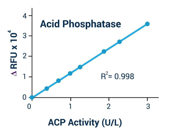
Analytical sensitivity is 0.2 U/L ACP, determined by adding two standard deviations to the mean OD of the zero standard assayed 20 times and calculating the corresponding concentration. The detection range is 0.2-50 U/L. Intra-assay precision across three human serum samples assayed in replicates of 20 ranged from 2.6% to 3.2% CV, with mean values of 0.50, 15.00, and 38.00 U/L. Inter-assay precision across 20 runs in duplicate by three operators ranged from 4.3% to 4.7% CV for the same samples.
| Product name: | Acid Phosphatase (ACP) Activity Assay Kit |
| SKU: | MAES0024 |
| Size: | 96 Assays |
| Sample type: | Serum, plasma, tissue |
| Detection method: | Colorimetric (PNPP substrate) |
| Detection instrument: | Microplate reader (400–415 nm; 405 nm optimum) |
| Assay time: | 30 min |
| Research area: | Enzymes; liver and renal function |
| Sensitivity: | 0.2 U/L |
| Detection range: | 0.2–50 U/L |
| Intra-assay CV: | 2.8% |
| Inter-assay CV: | 4.5% |
| Detection wavelength: | 405 nm |
| Sample volume: | 80 µL |
| Assay time: | 30 min |
- Colorimetric PNPP method read at 405 nm (400–415 nm microplate reader range)
- Detection range 0.2–50 U/L with analytical sensitivity of 0.2 U/L
- Validated for serum, plasma, and tissue homogenate samples
- Intra-assay CV 2.8% and inter-assay CV 4.5%
- Total assay time of 30 min; 96 assays per kit
Disodium p-nitrophenyl phosphate (PNPP), a widely used phosphatase chromogenic substrate, is converted to p-nitrophenol by acid phosphatase. Under alkaline conditions, p-nitrophenol is yellow with a maximum absorbance at 405 nm. Color intensity is proportional to ACP activity, so activity is calculated from the OD405 signal against a p-nitrophenol standard curve.
- Quantifying ACP activity in serum and plasma samples
- Measuring ACP activity in animal tissue homogenates normalized to protein concentration
- Comparing ACP activity across tissues in liver and renal function research
- Studying lysosomal acid hydrolase activity in cell and tissue preparations
- Profiling ACP activity in plant tissue extracts after pretest dilution optimization
| Component | Quantity | Storage |
| Reagent 1: Buffer Solution | 20 mL × 1 vial | -20°C, 12 months |
| Reagent 2: Substrate Powder | 3 vials | -20°C, 12 months, protect from light |
| Reagent 3: Standard Powder (p-nitrophenol) | 1 vial | -20°C, 12 months, protect from light |
| Reagent 4: Chromogenic Agent | 24 mL × 1 vial | -20°C, 12 months |
| Microplate | 1 plate (96 wells) | No requirement |
| Plate sealer | 2 pieces | No requirement |
| Sample layout sheet | 1 sheet | No requirement |
- Micropipettor and tips
- Microplate reader capable of 400–415 nm reading (405 nm optimum)
- 37°C incubator
- Double-distilled water
- PBS (0.01 M, pH 7.4)
- For tissue samples: dounce homogenizer, refrigerated centrifuge capable of 10000×g, cold PBS, and a user-supplied protein assay for protein concentration determination
Serum/plasma
- Detect serum or plasma directly.
- If not detected on the same day, store serum or plasma at -80°C for up to one month.
- Dilute samples before the assay. Recommended dilution factors are 5-10 for mouse plasma, rat plasma, and human plasma.
- Use normal saline (0.9% NaCl) or PBS (0.01 M, pH 7.4) as the diluent.
- For sample types not listed, perform a pretest to confirm the dilution factor.
Human urine
- Use human urine at a dilution factor of 1 (undiluted).
- For sample types not listed, perform a pretest to confirm the dilution factor.
Animal tissue
- Harvest the amount of tissue needed for each assay (initial recommendation 20 mg).
- Wash tissue in cold PBS (0.01 M, pH 7.4).
- Homogenize 20 mg tissue in 180 µL PBS (0.01 M, pH 7.4) with a dounce homogenizer at 4°C to yield a 10% homogenate.
- Centrifuge at 10000×g for 10 min at 4°C to remove insoluble material.
- Collect the supernatant and keep it on ice for detection.
- Determine the protein concentration of the supernatant with a separate protein assay, as required for the tissue calculation.
- Dilution factors for 10% homogenates: 20-30 for rat spleen, rat liver, and rat kidney tissue.
- Diluent is PBS (0.01 M, pH 7.4). For other tissue types, perform a pretest to confirm the dilution factor.
Plant tissue
- Prepare a 10% tissue homogenate. The listed example is 10% Epipremnum aureum tissue homogenate.
- Dilute the homogenate at a dilution factor of 5-10.
- Diluent is PBS (0.01 M, pH 7.4). For other plant tissue types, perform a pretest to confirm the dilution factor.
Reagent preparation
- Equilibrate all reagents to room temperature before use.
- Substrate working solution: dissolve one vial of Substrate Powder in 1.6 mL of Buffer Solution. Protect from light and use within 1 day.
- Standard stock (10 mmol/L): dissolve one vial of Standard Powder in 5 mL of double-distilled water and mix well. Aliquot and store at -20°C for up to 7 days, protected from light.
- Working 0.5 mmol/L standard: dilute 36 µL of standard stock with 684 µL of Buffer Solution. Prepare fresh on the day of use.
- Prepare the standard curve fresh for each assay and discard working dilutions after use.
Standard curve preparation (0.5 mmol/L standard and Buffer Solution)
- 0 mmol/L: 0 µL standard + 200 µL buffer
- 0.05 mmol/L: 20 µL standard + 180 µL buffer
- 0.1 mmol/L: 40 µL standard + 160 µL buffer
- 0.2 mmol/L: 80 µL standard + 120 µL buffer
- 0.25 mmol/L: 100 µL standard + 100 µL buffer
- 0.3 mmol/L: 120 µL standard + 80 µL buffer
- 0.4 mmol/L: 160 µL standard + 40 µL buffer
- 0.5 mmol/L: 200 µL standard + 0 µL buffer
Sample preparation
- Serum and plasma: assay directly. If not assayed the same day, store at -80°C for up to 1 month.
- Tissue: harvest the amount needed per assay (initial recommendation 20 mg) and wash in cold PBS (0.01 M, pH 7.4).
- Homogenize 20 mg tissue in 180 µL cold PBS (0.01 M, pH 7.4) with a dounce homogenizer at 4°C.
- Centrifuge at 10000×g for 10 min at 4°C. Collect the supernatant and keep on ice.
- Determine the protein concentration of the supernatant with a user-supplied protein assay.
- Dilute samples as needed. Reference dilution factors: mouse plasma 5–10; rat plasma 5–10; human plasma 5–10; human urine 1; 10% rat liver, spleen, or kidney homogenate 20–30. Dilute in PBS (0.01 M, pH 7.4). Pretest other sample types.
Assay procedure
- Add 40 µL of each standard to the standard wells, 40 µL of sample to the sample wells, and 40 µL of sample to the control wells.
- Add 40 µL of Buffer Solution to the standard wells and control wells.
- Add 40 µL of substrate working solution to the sample wells.
- Mix for 3 s on the microplate reader, then incubate at 37°C for 10 min.
- Add 160 µL of Chromogenic Agent to each well.
- Mix for 3 s on the microplate reader and read OD values at 405 nm.
Serum (plasma): ACP activity (U/L) = (ΔA - b) ÷ a ÷ T × f × 1000* Tissue: ACP activity (U/g protein) = (ΔA - b) ÷ a ÷ T × f × 1000* ÷ C_pr
- ΔA is the absorbed OD value, calculated as OD405 sample minus OD405 control.
- a is the slope and b is the intercept of the standard curve (y = ax + b), where y is the absorbed OD of the standard and x is its concentration in mmol/L.
- T is the reaction time, 10 min.
- f is the dilution factor of the sample before testing. Multiply by f to correct for sample dilution.
- C_pr is the protein concentration of the sample in g prot/L. Divide by C_pr to express tissue activity per gram of protein.
- 1000* converts units: 1 mmol = 1000 µmol. The serum result is in U/L, where 1 unit is the amount of enzyme that produces 1 µmol p-nitrophenol per liter of serum (plasma) per minute at 37°C.
- Example from the manual: for 10% rat kidney tissue homogenate diluted 20-fold with a protein concentration of 9.47 g prot/L, the reported result is 40.4 U/g prot.
What sample types can I test with this kit?
The kit measures ACP activity in serum, plasma, and tissue samples. The product page lists animal tissue, plant tissue, serum, and plasma, and the manual includes human urine dilution guidance.
How should samples and reagents be stored?
Reagents are stored at -20°C for 12 months, protected from light. Serum and plasma can be stored at -80°C for one month if not tested the same day, and substrate working solution should be used within one day.
What should I do if my sample is outside the detection range?
The detection range is 0.2-50 U/L. If the concentration is not within the detection range, take an extra dilution or concentration of the sample.
What are the key points for a reliable assay?
Protect substrate working solution and standards from light, and prepare a fresh set of standards for each run, discarding working dilutions after use. Equilibrate all reagents to room temperature before use and centrifuge small volumes before use.
What instrument settings are needed?
Use a microplate reader with a 400-415 nm filter range and an optimum wavelength of 405 nm. Mix each plate for 3 s before incubation and after adding chromogenic agent, then read OD at 405 nm. Incubate at 37°C.
What is the assay time?
The listed assay time is 30 min. The protocol includes a 10 min incubation at 37°C after substrate addition.