The RAP1A + RAP1B Monoclonal Antibody (CAB9725) is a high-quality antibody developed for reliable detection and analysis of target proteins. This antibody, generated in rabbits, exhibits high reactivity with human samples and has been validated for use in Western blot applications. By binding specifically to RAP1A and RAP1B proteins, researchers can accurately detect and analyze these critical signaling molecules in various cell types, making it a versatile option for studies in cell biology and cancer research.RAP1A and RAP1B are key regulators of cell adhesion and migration, influencing processes such as cell spreading, cell-cell adhesion, and cell-extracellular matrix interactions.
This antibody is validated for use in WB, IF/ICC, ELISA applications and has demonstrated reactivity against Human, Mouse, Rat samples.
Product Name:
RAP1A + RAP1B Monoclonal Antibody
SKU:
CAB9725
Size:
20μL, 100μL
Reactivity:
Human, Mouse, Rat
Clone Number:
ARC1719
Conjugate:
Unconjugated
Immunogen:
Synthetic peptide. This information is considered to be commercially sensitive.
This gene encodes a member of the RAS-like small GTP-binding protein superfamily. Members of this family regulate multiple cellular processes including cell adhesion and growth and differentiation. This protein localizes to cellular membranes and has been shown to regulate integrin-mediated cell signaling. This protein also plays a role in regulating outside-in signaling in platelets. Alternate splicing results in multiple transcript variants. Pseudogenes of this gene are found on chromosomes 3, 5, 6 and 9. [provided by RefSeq, Oct 2011]
Purification Method
Affinity purification
Gene ID
5908 5906
RRID
AB_2863766
Buffer Information
Store at -20℃. Avoid freeze / thaw cycles. Buffer: PBS containing 50% glycerol and 0.05% BSA, preserved with proclin300 or sodium azide, pH 7.3.
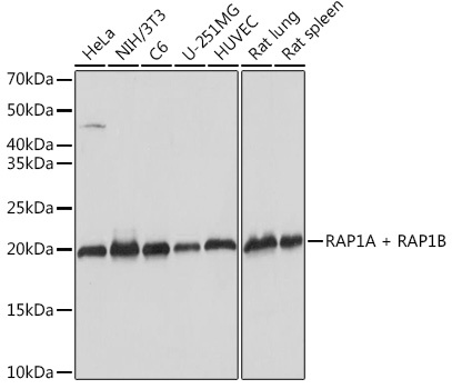
Western blot analysis of various lysates using RAP1A + RAP1B Rabbit mAb (CAB9725) at 1:1000 dilution. Secondary antibody: HRP-conjugated Goat anti-Rabbit IgG (H+L) (CABS014) at 1:10000 dilution. Lysates/proteins: 25μg per lane. Blocking buffer: 3% nonfat dry milk in TBST. Detection: ECL Basic Kit (AbGn00020). Exposure time: 1s.
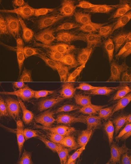
Immunofluorescence analysis of C6 cells using RAP1A + RAP1B Rabbit mAb (CAB9725) at dilution of 1:100 (40x lens). Secondary antibody: Cy3-conjugated Goat anti-Rabbit IgG (H+L) (CABS007) at 1:500 dilution. Blue: DAPI for nuclear staining.
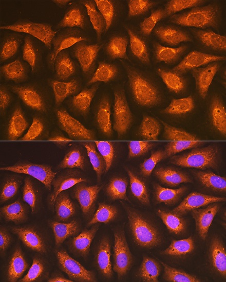
Immunofluorescence analysis of U-2 OS cells using RAP1A + RAP1B Rabbit mAb (CAB9725) at dilution of 1:100 (40x lens). Secondary antibody: Cy3-conjugated Goat anti-Rabbit IgG (H+L) (CABS007) at 1:500 dilution. Blue: DAPI for nuclear staining.


![Anti-RAP1A + RAP1B [R03-7H8] Monoclonal Antibody (AGMB01150)](https://cdn11.bigcommerce.com/s-h68l9z2lnx/images/stencil/590x590/products/272439/694501/anti-rap1a-rap1b-r03-7h8-monoclonal-antibody-agmb01150__40061.1774512682.jpg?c=2 "Anti-RAP1A + RAP1B [R03-7H8] Monoclonal Antibody (AGMB01150)")
![Anti-RAP1A+RAP1B [R03-1K4] Monoclonal Antibody (AGMB01151)](https://cdn11.bigcommerce.com/s-h68l9z2lnx/images/stencil/590x590/products/272440/692953/anti-rap1arap1b-r03-1k4-monoclonal-antibody-agmb01151__08951.1774507771.jpg?c=2 "Anti-RAP1A+RAP1B [R03-1K4] Monoclonal Antibody (AGMB01151)")


