Description
Calcein-AM/PI Double Staining Kit
The Assay Genie Calcein-AM/PI Double Staining Kit (AKES094) labels living and dead cells in one step. Calcein AM is converted by intracellular esterase to green-fluorescent Calcein in live cells, while propidium iodide (PI) enters cells with lost membrane integrity and binds double-stranded DNA to give red fluorescence. Researchers use the kit to assess cell viability and cytotoxicity by flow cytometry or fluorescence microscopy.
The Calcein AM/PI Double Staining Kit is a two-dye fluorescent system for distinguishing living and dead cells in mammalian cell populations. Calcein AM is the acetoxymethyl (AM) ester of calcein. The AM group makes the molecule hydrophobic enough to cross the intact plasma membrane, and the compound itself is not fluorescent. Its biological relevance depends on intracellular esterase activity, which converts it to a fluorescent readout only in cells that retain esterase function. Propidium iodide (PI) is the second component and marks cells that have lost selective membrane permeability.
The kit is intended for detecting cell activity and cytotoxicity. Because the two dyes are applied together, a single stained population can be scored for green-fluorescent living cells and red-fluorescent dead cells. The manual also describes treating cells with 5% to 20% DMSO for 2 to 4 hours or with 70% alcohol for 30 minutes to generate positive quality control samples. These controls allow the dead-cell signal to be verified before experimental samples are interpreted.
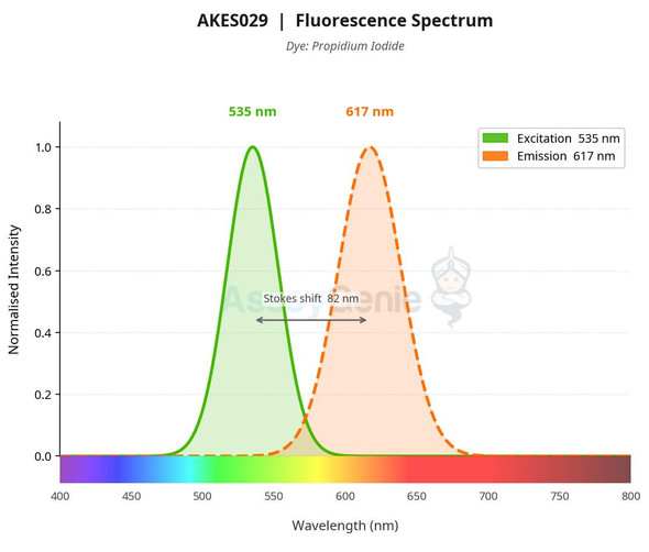
Calcein AM is hydrolyzed by endogenous esterases to calcein, a polar molecule with a strong negative charge that is retained inside living cells and emits green fluorescence (Ex/Em = 494 nm/517 nm). Dead cells lack esterase activity and produce little or no calcein. PI enters cells that have lost membrane integrity, binds double-stranded DNA, and emits red fluorescence (Ex/Em = 535 nm/617 nm). Cells are incubated with the combined working solution in the dark, then read by flow cytometry or fluorescence microscopy.
The kit is designed for animal cells that contain esterase activity. The manual states that it is not suitable for plant or bacterial cells because Calcein AM cannot enter the cell wall. Cells should be washed with serum-free medium or PBS before staining, because serum may contain esterase. The staining buffer must not contain primary or secondary amines, which can lyse AM esters and hinder dye loading. Metal ions such as Mn2+ quench fluorescence and should be excluded from wash buffers. Calcein AM Solution should be packaged and protected from light before its first use to prevent spontaneous hydrolysis in a damp environment.
| Detection method: | Fluorometric; flow cytometry |
| Sample type: | Cells (animal cells with esterase activity) |
| Assay time: | 30 min |
| Detection instrument: | Flow cytometer; fluorescence microscope |
| Dye type: | Calcein/PI |
| Excitation/emission (nm): | Calcein 494/517; PI 535/617 |
| Flow cytometry channels: | Calcein: FITC; PI: PE or PerCP/Cy5.5 |
| Microscopy filter set: | FITC, TRITC |
- Dual live/dead staining: green Calcein (Ex/Em 494/517 nm) for live cells, red PI (Ex/Em 535/617 nm) for dead cells
- Compatible with flow cytometry and fluorescence microscopy
- Staining time of approximately 30 min
- Suitable for animal cells with esterase activity
- Calcein AM Solution (100 µmol/L), PI Solution (750 µmol/L) and Calcein AM Assay Buffer supplied
- Research use only
Calcein AM passes through intact membranes and is hydrolyzed by endogenous esterase to calcein, which is retained in live cells and fluoresces green (Ex/Em 494/517 nm). Dead cells have lost esterase activity and membrane integrity, so they show little or no calcein signal and instead take up PI, which fluoresces red (Ex/Em 535/617 nm) on binding double-stranded DNA.
- Live/dead discrimination of cell populations by flow cytometry
- Cell viability assessment by fluorescence microscopy
- Cytotoxicity evaluation of compound- or drug-treated cells
- Single-stain compensation controls for multicolor flow cytometry panels
| Component | Storage |
| Calcein AM Solution (100 µmol/L) | -20°C, protected from light |
| PI Solution (750 µmol/L) | 2–8°C or -20°C, protected from light |
| Calcein AM Assay Buffer | -20°C |
- Phosphate-buffered saline (PBS), pH 7.2–7.4
- Flow cytometer or fluorescence microscope with FITC and PE or PerCP/Cy5.5 (flow) or FITC and TRITC (microscopy) filter sets
- Centrifuge capable of 300×g
- Vortex mixer
- Water bath or room temperature bench for thawing
- 37°C incubator (microscopy protocol)
- Serum-free medium or PBS for washing cells
- Cell suspension or adherent cells in 96-well or 24-well plates
Cells in suspension (flow cytometry)
- Collect cells and centrifuge at 300 × g for 5 min. Discard the supernatant.
- Resuspend the cell pellet in 1 mL of PBS, centrifuge at 300 × g for 5 min, and discard the supernatant. Wash once.
- Prepare the staining working solution fresh before use. For 1 mL, combine 0.1 µL of Calcein AM Solution (100 µM), 1 µL of PI Solution (750 µM), and 1 mL of Calcein AM Assay Buffer.
- For greater reagent economy, dilute Calcein AM Solution (100 µM) 100-fold to 1 µM with Calcein AM Assay Buffer. Then add 2 µL of the 1 µM solution to 200 µL of Calcein AM Assay Buffer for staining.
- Add 200 µL of staining working solution to resuspend 1 to 5 × 10^5 cells per sample. Incubate for 5 to 15 min at room temperature in the dark.
- Analyze immediately by flow cytometry. If analysis is delayed, keep samples protected from light at 4°C and detect within 2 hours.
- Prepare negative control and single-stain tubes with the single-dye working solutions described in the manual (Calcein AM alone, PI alone, and buffer alone) for compensation adjustment.
Adherent cells (fluorescence microscopy)
- Remove the culture medium carefully. Add an appropriate volume of PBS to each well to wash the cells, then repeat the wash and remove the PBS.
- Prepare the staining working solution fresh. For 1 mL, combine 10 µL of Calcein AM Solution (10 µM), 10 µL of PI Solution (750 µM), and 1 mL of Calcein AM Assay Buffer. Scale volumes to the number of samples.
- Add 100 µL of working solution per well in a 96-well plate or 200 µL per well in a 24-well plate.
- Incubate at 37°C for 10 to 30 min. If the working solution is prepared with basic medium, extend the incubation to 30 to 60 min and add PI 10 min before the end of the reaction.
- Observe under a fluorescence microscope. Calcein appears green, and PI appears red.
Suspended cells (fluorescence microscopy)
- After collecting the cell pellet, add staining working solution at 1 to 5 × 10^5 cells per 200 µL.
- Incubate at room temperature for 15 to 20 min.
- Place the cell suspension on a glass slide, cover gently with a coverslip, and observe under the microscope.
Positive control cells
- Treat cells with 5% to 20% DMSO for 2 to 4 hours, or with 70% alcohol for 30 min, to obtain positive quality control samples for dead-cell staining.
Flow cytometry
- Thaw the kit reagents at room temperature and vortex each reagent before use.
- Prepare the staining working solution fresh: for 1 mL, combine 0.1 µL Calcein AM Solution (100 µmol/L), 1 µL PI Solution (750 µmol/L) and 1 mL Calcein AM Assay Buffer. Scale volumes proportionally for 5 mL and 10 mL preparations.
- Prepare single-stain and negative control tubes as needed: Calcein AM single stain (0.1 µL Calcein AM Solution in 1 mL buffer), PI single stain (1 µL PI Solution in 1 mL buffer), and unstained negative control (1 mL buffer only).
- Collect cells, centrifuge at 300×g for 5 min and discard the supernatant. Resuspend in 1 mL PBS, centrifuge again at 300×g for 5 min and discard the supernatant.
- Resuspend 1–5×10⁵ cells in 200 µL of staining working solution and incubate for 5–15 min at room temperature in the dark.
- Analyze directly by flow cytometry. If analysis is delayed, protect from light and store at 4°C for up to 2 hours before detection.
Fluorescence microscopy
- Thaw reagents at room temperature and vortex each reagent before use.
- Prepare the staining working solution fresh per well: 100 µL per well of a 96-well plate or 200 µL per well of a 24-well plate. For 1 mL, combine 10 µL of the Calcein AM solution, 10 µL PI Solution (750 µmol/L) and 1 mL Calcein AM Assay Buffer, as listed in the reagent table for this protocol.
- Remove culture medium from adherent cells, wash with PBS and remove the PBS.
- Add staining working solution (100 µL per well in 96-well plates or 200 µL per well in 24-well plates) and incubate at 37°C for 10–30 min. Add PI 10 min before the end of incubation when using basic medium-based working solution, which requires a 30–60 min staining time.
- Observe under a fluorescence microscope: Calcein green (Ex/Em 494/517 nm), PI red (Ex/Em 535/617 nm).
- For suspended cells, collect the cell pellet and resuspend at 1–5×10⁵ cells per 200 µL of working solution. Incubate at room temperature for 15–20 min, mount on a glass slide and cover gently before observation.
Which sample types are compatible with this kit?
The kit is intended for mammalian and other animal cells that have esterase activity. Plant and bacterial cells are not suitable because Calcein AM cannot enter the cell wall.
How should the kit and working solutions be stored?
The kit should be stored at -20°C, protected from light, for 12 months. Calcein AM Solution should be packaged and protected from light before first use. Prepare the Calcein AM/PI working solution fresh before each use, and keep flow cytometry samples protected from light at 4°C if analysis is delayed, detecting them within 2 hours.
What should I do if PI-positive signal appears in cells that should be viable?
The manual warns that the PI staining time should be less than 30 min, otherwise false-positive PI signal may occur. If Calcein AM staining must be extended, add PI within 10 to 30 min before the end of Calcein AM staining, and observe and image within 1 to 2 hours.
What factors can interfere with staining?
Wash cells with serum-free medium or PBS, because serum may contain esterase. Do not use buffers containing primary or secondary amines, which can lyse AM esters and hinder loading. Avoid metal ions such as Mn2+, which quench fluorescence. Calcein is also excluded by P-glycoprotein multidrug transporters.
Which instrument channels and filters should I use?
For flow cytometry, detect Calcein in the FITC channel and PI in the PE or PerCP/Cy5.5 channel. For fluorescence microscopy, use FITC and TRITC filter sets. Calcein is excited at 494 nm and emits at 517 nm, and PI is excited at 535 nm and emits at 617 nm.
How long does the assay take?
The product page lists an assay time of 30 min. Flow cytometry staining requires 5 to 15 min at room temperature in the dark. Microscopy staining requires 10 to 30 min at 37°C, or 30 to 60 min when basic medium is used for the working solution.
| Storage: | -20°C for 12 months, protected from light |
| Shelf life: | 12 months |
| Shipping: | Ice bag |
| Handling: | Package and store Calcein AM Solution protected from light before first use to prevent spontaneous ester hydrolysis in a humid environment |
Cautions
- For research use only. Not for use in diagnostic or therapeutic procedures.
- Wash cells with serum-free medium or PBS before staining; serum may contain esterase.
- Do not use buffers containing primary or secondary amines, which can hydrolyze AM esters and hinder dye loading.
- Avoid metal ions such as Mn2+ in wash buffers; Mn2+ quenches fluorescence.
- Calcein AM working solution is hygroscopic; prepare fresh before use.
- Staining at 37°C shortens staining time; staining at room temperature reduces dye penetration into organelles.
- Calcein AM is not suitable for plant cells or bacteria.
- Excessive centrifuge acceleration or deceleration may cause cell loss; use acceleration no higher than 3 and deceleration no higher than 2.
- For microscopy of weakly adherent cells, consider anti-detachment treatment of culture vessels. Keep PI staining under 30 min to avoid false-positive PI signal.
- Positive controls can be generated by treating cells with 5%–20% DMSO for 2–4 hours or 70% alcohol for 30 min.