Description
Fructose Fluorometric Assay Kit
The Fructose Fluorometric Assay Kit (MAES0289) quantifies fructose in serum, plasma, and tissue homogenate samples. The enzymatic reaction produces a product that reacts with a chromogenic agent to generate a fluorescent signal read at Ex/Em 530/590 nm. Researchers use the kit to study fructose levels in metabolic disease models and in fruit and plant tissues.
Fructose is a ketohexose, the most common ketose found in nature. It occurs in large amounts as a free monosaccharide in fruits and honey, and it is a dietary sugar that is metabolized differently from glucose. Its concentration in biological and food samples is a useful indicator of carbohydrate content, which makes accurate quantification a routine need in metabolic and plant studies.
Researchers measure fructose because large epidemiological datasets and experimental studies associate excessive fructose intake with an increased incidence of metabolic diseases. The kit is designated for glycolysis and lipid metabolism research, and it is also used in plant physiology, where fruit sugar content is a key measurement. Tissue homogenates from watermelon, cantaloupe, orange, mango, grape, and pineapple are specifically addressed, along with mouse liver tissue and mouse, human, and rabbit serum.
The assay is enzymatic. Fructose is converted by enzymes into a specific product, which then reacts with a chromogenic agent to form a fluorescent substance. The workflow adds enzyme working solution to each well and incubates at 37 °C for 90 min protected from light. Chromogenic working solution is then added to standard and sample wells, and substrate working solution is added to control wells. After a further 30 min incubation at 37 °C, fluorescence is read at an excitation of 530 nm and an emission of 590 nm on a fluorescence microplate reader, using a black 96-well plate.
Serum and plasma can be assayed directly. If they are not tested on the same day, they can be stored at -80 °C for up to one month. Tissue is washed in cold PBS (0.01 M, pH 7.4), homogenized at 4 °C in normal saline (0.9% NaCl) at a ratio of 20 mg tissue to 180 µL diluent, and centrifuged at 10000 × g for 10 min at 4 °C. The supernatant is kept on ice for testing. Because tissue results are normalized to protein, the protein concentration of each supernatant must be measured separately. Other sample types require a pretest to confirm dilution.
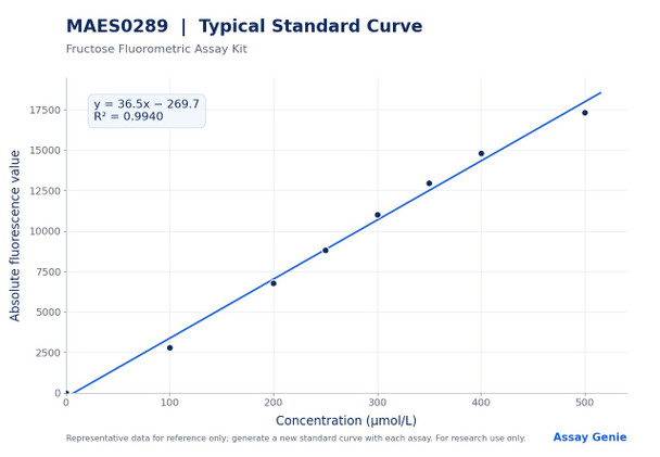
Sensitivity is 0.72 µmol/L, and the detection range is 0.72 to 500 µmol/L. Intra-assay precision is 1.7 to 3.1% CV, and inter-assay precision is 3.2 to 7.3% CV. Spike-recovery experiments at low, middle, and high concentrations gave individual recoveries of 95 to 111%, with an average recovery of 101%. The standard curve values are provided for reference only, because fluorescence can vary with operator, pipetting technique, and temperature. Each run should therefore include its own freshly prepared standards.
| Product Name: | Fructose Fluorometric Assay Kit |
| SKU: | MAES0289 |
| Sample Type: | Serum, plasma, tissue |
| Assay Type: | Quantitative |
| Detection Method: | Fluorometric (enzymatic) |
| Detection Instrument: | Fluorescence microplate reader (Ex/Em 530 nm/590 nm) |
| Assay Time: | 2 h 30 min |
| Research Area: | Glycolysis and lipid metabolism; plant physiology |
| Sensitivity: | 0.72 µmol/L |
| Detection Range: | 0.72-500 µmol/L |
| Sample Volume per Well: | 20 µL |
| Intra-assay CV (50 µmol/L, 150 µmol/L, 350 µmol/L): | 1.7%, 2.3%, 3.1% |
| Inter-assay CV (50 µmol/L, 150 µmol/L, 350 µmol/L): | 3.2%, 5.2%, 7.3% |
| Average Recovery: | 101% |
| Recovery Rate (50 µmol/L, 150 µmol/L, 350 µmol/L): | 97%, 95%, 111% |
- Quantitative fluorometric detection of fructose, read at Ex/Em 530 nm/590 nm
- Detection range 0.72-500 µmol/L with analytical sensitivity of 0.72 µmol/L
- Validated for serum, plasma, and tissue homogenate samples
- Total assay time of 2 h 30 min, including 90 min and 30 min incubations at 37°C
- Intra-assay CV 1.7-3.1% and inter-assay CV 3.2-7.3% across three sample concentrations

Fructose is converted by enzymes into a specific product. This product reacts with the chromogenic agent to form a fluorescent substance, which is measured at Ex/Em 530 nm/590 nm. Fluorescence in each sample is corrected against a control well, and fructose concentration is calculated from a standard curve.
- Quantification of fructose in serum and plasma for metabolic disease research
- Measurement of fructose in tissue homogenates from animal models
- Analysis of fructose content in fruit and plant tissue samples for plant physiology studies
- Investigation of fructose's role in glycolysis and lipid metabolism
| Component | Quantity | Storage |
| Reagent 1, Buffer Solution | 50 mL × 1 vial | -20°C, 12 months |
| Reagent 2, Matrix Solution | 50 mL × 1 vial | -20°C, 12 months; protect from light |
| Reagent 3, Enzyme Reagent | Powder × 8 vials | -20°C, 12 months; protect from light |
| Reagent 4, Chromogenic Agent | Powder × 8 vials | -20°C, 12 months; protect from light |
| Reagent 5, Substrate | Powder × 8 vials | -20°C, 12 months; protect from light |
| Reagent 6, Accelerant | 1 mL × 1 vial | Protect from light |
| Reagent 7, 5 mmol/L Standard Solution | 1.6 mL × 1 vial | -20°C, 12 months |
| Black microplate | 96 wells | Room temperature |
| Plate sealer | 2 pieces | Room temperature |
| Sample layout sheet | 1 piece | Room temperature |
- Fluorescence microplate reader (Ex/Em 530 nm/590 nm)
- Incubator set to 37°C
- Vortex mixer
- Normal saline (0.9% NaCl)
- Double-distilled water (for standard dilutions)
- PBS (0.01 M, pH 7.4) for tissue washing
- Homogenizer and refrigerated centrifuge capable of 10000 × g
- Protein assay reagents for tissue normalization
Serum/plasma
- Detect serum or plasma directly.
- If samples are not tested on the same day, store serum or plasma at -80 °C for up to one month.
- Recommended dilution factor: 1 for mouse serum, human serum, and rabbit serum.
- Dilute with normal saline (0.9% NaCl) or PBS (0.01 M, pH 7.4).
- For other sample types, perform a pretest to confirm the dilution factor.
Tissue
- Harvest the amount of tissue needed for each assay (initial recommendation: 20 mg).
- Wash the tissue in cold PBS (0.01 M, pH 7.4).
- Homogenize 20 mg tissue in 180 µL normal saline (0.9% NaCl) with a dounce homogenizer at 4 °C.
- Centrifuge at 10000 × g for 10 min at 4 °C to remove insoluble material. Collect the supernatant and keep it on ice for detection.
- Determine the protein concentration of the supernatant using a separate protein assay.
- Recommended dilution factors (for reference only): 10% watermelon tissue homogenate 40–90; 10% cantaloupe tissue homogenate 30–90; 10% orange tissue homogenate 20–80; 10% mango tissue homogenate 30–90; 10% grape tissue homogenate 60–200; 10% pineapple tissue homogenate 15–40; 10% mouse liver tissue homogenate 1.
- Diluent: normal saline (0.9% NaCl) or PBS (0.01 M, pH 7.4). For other sample types, perform a pretest to confirm the dilution factor.
Reagent preparation
- Equilibrate all reagents to room temperature before use.
- Enzyme working solution: dissolve one vial of Reagent 3 in 1.125 mL of Reagent 1, mix well, then add 2 µL of Reagent 6 and mix. Store at 2-8°C for up to 1 week, protected from light.
- Chromogenic working solution: dissolve one vial of Reagent 4 in 1.5 mL of Reagent 2. Store at 2-8°C for up to 3 days, protected from light.
- Substrate working solution: dissolve one vial of Reagent 5 in 1.5 mL of Reagent 2. Store at 2-8°C for up to 3 days, protected from light.
- 500 µmol/L standard solution: mix 100 µL of Reagent 7 (5 mmol/L) with 900 µL of double-distilled water. Store at 2-8°C for up to 1 week, protected from light.
Standard curve preparation
- Prepare a fresh set of standards before each assay and discard working dilutions after use.
- Dilute the 500 µmol/L standard with double-distilled water to 0, 100, 200, 250, 300, 350, 400, and 500 µmol/L (for example, 0, 40, 80, 100, 120, 140, 160, and 200 µL of 500 µmol/L standard, brought to 200 µL total with 200, 160, 120, 100, 80, 60, 40, and 0 µL of water).
Sample preparation
- Serum and plasma: assay directly. If not assayed the same day, store at -80°C for up to one month.
- Tissue: weigh out tissue for each assay (initial recommendation 20 mg) and wash in cold PBS (0.01 M, pH 7.4).
- Homogenize 20 mg of tissue in 180 µL of normal saline (0.9% NaCl) on ice.
- Centrifuge at 10000 × g for 10 min at 4°C. Collect the supernatant and keep it on ice for detection.
- Determine supernatant protein concentration for tissue normalization.
- Dilute serum or tissue samples as needed. Recommended dilution factors: human, mouse, and rabbit serum, 1; mouse liver homogenate, 1; 10% watermelon homogenate, 40-90; 10% cantaloupe homogenate, 30-90; 10% orange homogenate, 20-80; 10% mango homogenate, 30-90; 10% grape homogenate, 60-200; 10% pineapple homogenate, 15-40. Use normal saline (0.9% NaCl) or PBS (0.01 M, pH 7.4) as diluent.
Serum/plasma: Fructose content (µmol/L) = (ΔF - b) / a × f Tissue: Fructose content (mmol/gprot) = (ΔF - b) / a ÷ Cpr ÷ 1000 × f
- ΔF is the absolute fluorescence value of the sample, calculated as F sample - F control.
- a is the slope of the standard curve, and b is the intercept of the standard curve (standard curve: y = ax + b).
- The absolute fluorescence value of each standard is obtained by subtracting the mean fluorescence of the blank (standard ①, 0 µmol/L) from all standard readings. The standard curve plots absolute fluorescence (y-axis) against concentration in µmol/L (x-axis).
- f is the dilution factor of the sample before testing.
- Cpr is the protein concentration of the tissue sample, in gprot/L.
- 1000 is the conversion factor, where 1 mmol = 1000 µmol.
- Serum/plasma results are reported in µmol/L. Tissue results are reported in mmol/gprot.
What sample types can be tested with this kit?
The kit measures fructose in serum, plasma, and tissue samples. Tissue homogenates from watermelon, cantaloupe, orange, mango, grape, and pineapple are listed with recommended dilution factors, as are mouse liver tissue and mouse, human, and rabbit serum.
How should samples and reagents be stored?
Kit components should be stored at -20 °C for 12 months, with the enzyme, chromogenic agent, and substrate protected from light. Serum and plasma not tested on the same day can be stored at -80 °C for up to one month. Working solutions should be stored at 2–8 °C protected from light: enzyme working solution for 1 week, and chromogenic and substrate working solutions for 3 days.
What is the detection range, and what should I do if my sample is out of range?
The detection range is 0.72 to 500 µmol/L. If the concentration falls outside this range, dilute or concentrate the sample, and apply the appropriate dilution factor in the calculation.
What interferences or key points should I observe?
Use the kit for research use only and not for clinical diagnosis. Protect the enzyme, chromogenic agent, and substrate from light, and do not mix reagents from different kits. Prepare a fresh set of standards for each assay and discard working standard dilutions after use.
What instrument settings and assay time are required?
Read fluorescence on a fluorescence microplate reader at an excitation of 530 nm and an emission of 590 nm, using a black 96-well plate. Each well receives 20 µL of standard or sample. The total assay time is 2 h 30 min, including the 90 min and 30 min incubations at 37 °C.
| Storage: | -20°C, protect from light |
| Shelf Life: | 12 months |
Notes
- For research use only; do not use in clinical diagnosis.
- Store reagents strictly according to the preservation conditions; do not mix reagents from different kits.
- Equilibrate all reagents to room temperature before use.
- Protect enzyme, chromogenic, and substrate working solutions from light during storage and incubation.
- Centrifuge small-volume reagents before use to ensure sufficient recovery.
- Prepare fresh standard curve dilutions for each assay and discard working standards after use.