Description
GABA Colorimetric Assay Kit
The GABA Colorimetric Assay Kit (MAES0202) quantifies γ-aminobutyric acid (GABA) in animal and plant tissue homogenates. Phenol and sodium hypochlorite react with GABA to form a blue-green product read at 640 nm on a microplate reader. The kit is used in studies of GABA content in plant physiology and animal tissue neurochemistry.
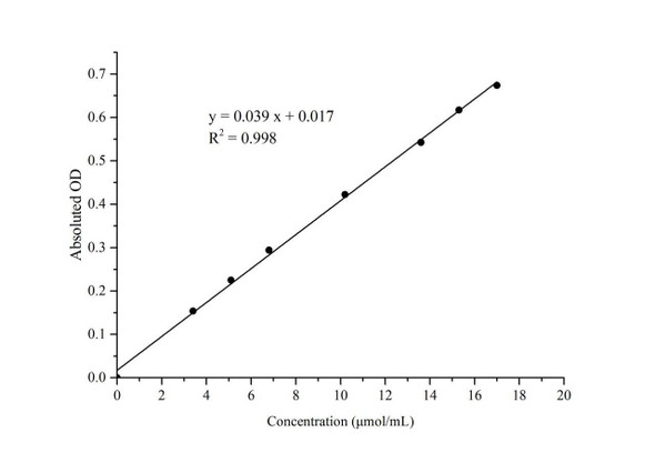
γ-Aminobutyric acid (GABA) is the analyte quantified by the GABA Colorimetric Assay Kit (MAES0202) in animal and plant tissue samples. Results are expressed as GABA content per gram of wet tissue weight. The kit includes a 10 µmol/mL GABA standard solution that is diluted with extracting solution to build a calibration curve spanning 0 to 10 µmol/mL. Standard dilutions are prepared fresh for each run and discarded after use.
Researchers use this kit to measure GABA content in animal and plant tissues. The product is indexed under Amino Acids and Proteins and Plant Physiology, reflecting its use in plant studies as well as animal work. The source materials list rat heart, rat liver, and rat kidney tissues, and plant tissues including Epipremnum aureum, green pepper, and Chinese yam, each with a recommended dilution factor, as examples of sample types the kit has been used with.
The assay is based on a reaction in which phenol and sodium hypochlorite react with GABA to produce a blue-green product with maximum absorbance at 640 nm. For each standard or sample, 30 µL is mixed with 50 µL buffer solution and 40 µL chromogenic agent A, vortexed, and held at room temperature for 5 min. Chromogenic agent B (60 µL) is then added, and tubes are vortexed, heated in a 95°C water bath for 10 min, and cooled in an ice bath. After adding 200 µL of supplementary solution, 200 µL from each tube is transferred to a microplate and read at 640 nm on a microplate reader set to 630-650 nm. Readings should be taken within 10 min after the reaction, and the total assay time is 40 min.
Suitable samples are animal tissue and plant tissue homogenates prepared as described in the sample preparation section. Tissue is washed in cold PBS (0.01 M, pH 7.4) and homogenized in extracting solution at 4°C, then heated and centrifuged before testing. Kit components should be stored at 2-8°C for 12 months, and the chromogenic agents must be kept away from light. Reagents from different kits must not be mixed, and small reagent volumes should be centrifuged before use to ensure sufficient recovery.
Performance data from the manual and product page show an analytical sensitivity of 0.06 µmol/mL, determined as two standard deviations above the mean OD of the zero standard across 20 replicates. The detection range is 0.06-10.0 µmol/mL. In rat heart tissue samples, intra-assay CV ranged from 3.8% to 4.3% and inter-assay CV ranged from 6.6% to 7.2%. Mean recovery across three spiked concentrations was 96%, with individual recoveries of 95% to 98%.
| Product Name: | GABA Colorimetric Assay Kit |
| SKU: | MAES0202 |
| Size: | 96 Assays |
| Sample Type: | Animal tissue, plant tissue |
| Detection Method: | Colorimetric |
| Instrument: | Microplate reader (630-650 nm; optimum 640 nm) |
| Assay Time: | 40 min |
| Research Area: | Amino acids and proteins; plant physiology |
| Use: | Research Use Only |
| Sensitivity: | 0.06 µmol/mL |
| Detection Range: | 0.06-10.0 µmol/mL |
| Average Recovery: | 96% |
| Intra-assay CV: | 4% |
| Inter-assay CV: | 6.8% |
| Sample Volume: | 30 µL tissue homogenate supernatant per well |
| Assay Time: | 40 min |
- Quantifies GABA in animal and plant tissue samples
- Colorimetric readout at 640 nm (microplate reader, 630-650 nm filter range)
- Detection range 0.06-10.0 µmol/mL; sensitivity 0.06 µmol/mL
- Assay time 40 min after tissue extraction
- Average recovery 96%; intra-assay CV 4%, inter-assay CV 6.8%
- Supplied as 96 assays with GABA standard included
Phenol and sodium hypochlorite react with GABA to produce a blue-green product with maximum absorbance at 640 nm. GABA content is calculated from the absorbance at 640 nm against a standard curve prepared with a 10 µmol/mL GABA standard.
- Measurement of GABA content in plant tissues such as Epipremnum aureum, green pepper, and Chinese yam homogenates
- Measurement of GABA content in rat heart, liver, and kidney tissue homogenates
- Plant physiology studies of non-protein amino acid accumulation
- Neurochemistry studies of GABA levels in animal tissue
- Comparative studies of GABA content across tissue types or treatment conditions
| Component | Quantity | Storage |
| Reagent 1 (Extracting Solution) | 60 mL × 2 vials | 2-8°C, 12 months |
| Reagent 2 (Buffer Solution) | 6 mL × 1 vial | 2-8°C, 12 months |
| Reagent 3 (Chromogenic Agent A) | 4.8 mL × 1 vial | 2-8°C, 12 months, shading light |
| Reagent 4 (Chromogenic Agent B) | 7.2 mL × 1 vial | 2-8°C, 12 months, shading light |
| Reagent 5 (Supplementary Solution) | 24 mL × 1 vial | 2-8°C, 12 months |
| Reagent 6 (10 µmol/mL GABA Standard) | 1.6 mL × 2 vials | 2-8°C, 12 months |
| Microplate | 96 wells | No requirement |
- Test tubes (1.5 mL EP tubes)
- Micropipettor
- Vortex mixer
- Microplate reader (630-650 nm; optimum 640 nm)
- Centrifuge (capable of 8000 × g)
- Water bath (95°C)
- Ice bath
- Homogenizer (dounce type)
- Cold PBS (0.01 M, pH 7.4) for tissue washing
- Ice or 4°C storage for homogenization
Tissue
- Harvest the amount of tissue needed for each assay (initial recommendation 50 mg).
- Wash tissue in cold PBS (0.01 M, pH 7.4).
- Homogenize 50 mg tissue in 450 µL extracting solution with a Dounce homogenizer at 4°C.
- After homogenizing, transfer to an EP tube and mark the liquid level.
- Heat in a 95°C water bath for 2 h. Keep the EP tube cover tight, and tie the tube cover with a small hole for ventilation to prevent the cover from bursting open under high temperature.
- Supplement the supernatant with extracting solution to the original volume and mix fully. Centrifuge at 8000 × g for 10 min and take the supernatant for detection.
- Dilution: the recommended dilution factor is 1 for 10% homogenates of Epipremnum aureum tissue, green pepper tissue, Chinese yam tissue, rat heart tissue, rat liver tissue, and rat kidney tissue. The diluent is extracting solution. For other sample types, perform a pretest to confirm the dilution factor.
Sample preparation (tissue)
- Harvest the tissue amount needed for each assay (initial recommendation 50 mg).
- Wash tissue in cold PBS (0.01 M, pH 7.4).
- Homogenize 50 mg tissue in 450 µL extracting solution with a dounce homogenizer at 4°C.
- Transfer to an EP tube and mark the liquid level. Heat in a 95°C water bath for 2 h with a loosely fitted cap (small vent hole in the cover).
- Supplement the supernatant with extracting solution to the original volume and mix. Centrifuge at 8000 × g for 10 min and collect the supernatant for detection.
- Dilution factor is 1 for the listed tissue homogenates (10% Epipremnum aureum, green pepper, Chinese yam, rat heart, rat liver, rat kidney). Diluent is extracting solution; pretest other sample types to confirm dilution.
Standard curve preparation (prepare fresh)
- Equilibrate all reagents to room temperature.
- Dilute the 10 µmol/mL GABA standard with extracting solution to 0, 1, 2, 4, 5, 7, 9, and 10 µmol/mL using: 0 µL standard/200 µL extracting solution (0); 20/180 µL (1); 40/160 µL (2); 80/120 µL (4); 100/100 µL (5); 140/60 µL (7); 180/20 µL (9); 200/0 µL (10).
- Discard working standard dilutions after use.
Assay
- Standard tubes: transfer 30 µL of each standard to 1.5 mL EP tubes. Sample tubes: transfer 30 µL of sample supernatant to 1.5 mL EP tubes.
- Add 50 µL Reagent 2 (buffer solution) and 40 µL Reagent 3 (Chromogenic Agent A) to each tube.
- Mix fully on a vortex mixer and stand at room temperature for 5 min.
- Add 60 µL Reagent 4 (Chromogenic Agent B) to each tube.
- Mix fully on a vortex mixer and heat in a 95°C water bath for 10 min. Cool in an ice bath.
- Add 200 µL Reagent 5 (supplementary solution) to each tube and mix fully.
- Transfer 200 µL from each tube to the microplate and read OD at 640 nm. Read within 10 min after the reaction.
Calculation
- Average duplicate readings for each standard.
- Subtract the mean OD of the blank (standard 0 µmol/mL) from all standard readings to obtain absolute OD.
- Plot absolute OD (y) against concentration (x) and fit y = ax + b.
- Calculate GABA content (µmol/g wet weight) = (ΔA640 − b) ÷ a × V ÷ m × f, where ΔA640 = OD sample − OD blank, V = extracting solution volume (mL), m = tissue weight (g), and f = dilution factor.
GABA content (µmol/g wet weight) = (ΔA640 - b) ÷ a × V ÷ m × f
- ΔA640 = OD(Sample) - OD(Blank), where OD(Blank) is the OD of the standard with concentration 0 µmol/mL. Absolute OD values of standards are calculated by subtracting the mean blank OD from all standard readings.
- a and b are the slope and intercept of the standard curve y = ax + b, where y is absolute OD and x is GABA concentration (µmol/mL).
- V is the volume of extracting solution in mL.
- m is the weight of tissue in g.
- f is the dilution factor of the sample before testing. Undiluted samples have f = 1.
- Example: for green pepper tissue with standard curve y = 0.034x + 0.0032, sample OD 0.135, blank OD 0.043, V = 0.9 mL, m = 0.1 g, and f = 1, the result is 23.49 µmol/g wet weight.
Which sample types are supported?
The kit measures GABA in animal tissue and plant tissue samples. Tissue is homogenized in extracting solution, heated at 95°C for 2 h, and centrifuged at 8000 × g for 10 min, and 30 µL of the supernatant is used per assay.
How should the kit and reagents be stored?
Kit components should be stored at 2-8°C for 12 months. The chromogenic agents must also be protected from light, and reagents from different kits must not be mixed.
What should I do if my sample is out of the detection range?
The detection range is 0.06-10.0 µmol/mL. If the GABA concentration is not within this range, the manual recommends an extra dilution or concentration of the sample before testing.
What key points affect the assay?
Equilibrate all reagents to room temperature before use and prepare a fresh set of standards for each run. Take readings within 10 min after the reaction, as the assay is designed to be determined within that window.
What instrument settings should I use?
Read the absorbance on a microplate reader set to 630-650 nm, with 640 nm as the optimum wavelength. Transfer 200 µL from each tube to the microplate for reading.
How long does the assay take?
The total assay time is 40 min.
| Storage: | 2-8°C |
| Shelf Life: | 12 months |
| Light Protection: | Chromogenic Agents A and B: store with shading light protection |
| Shipping: | Gel pack |
Notes
- For Research Use Only. Not for clinical diagnosis or therapeutic use.
- Read the instructions completely and adjust instruments before use.
- Wear a lab coat and latex gloves.
- Prepare a fresh standard curve for each assay and discard working dilutions after use.
- Do not mix reagents from different kits.
- Centrifuge small-volume reagents before use.
- If a sample falls outside the detection range, dilute or concentrate it and repeat the assay.
- Run a pretest for sample types not listed in the protocol.
- Determine results within 10 min after the reaction step.