Description
Mycoplasma detection kit for cell culture: a visual result in 1 hour
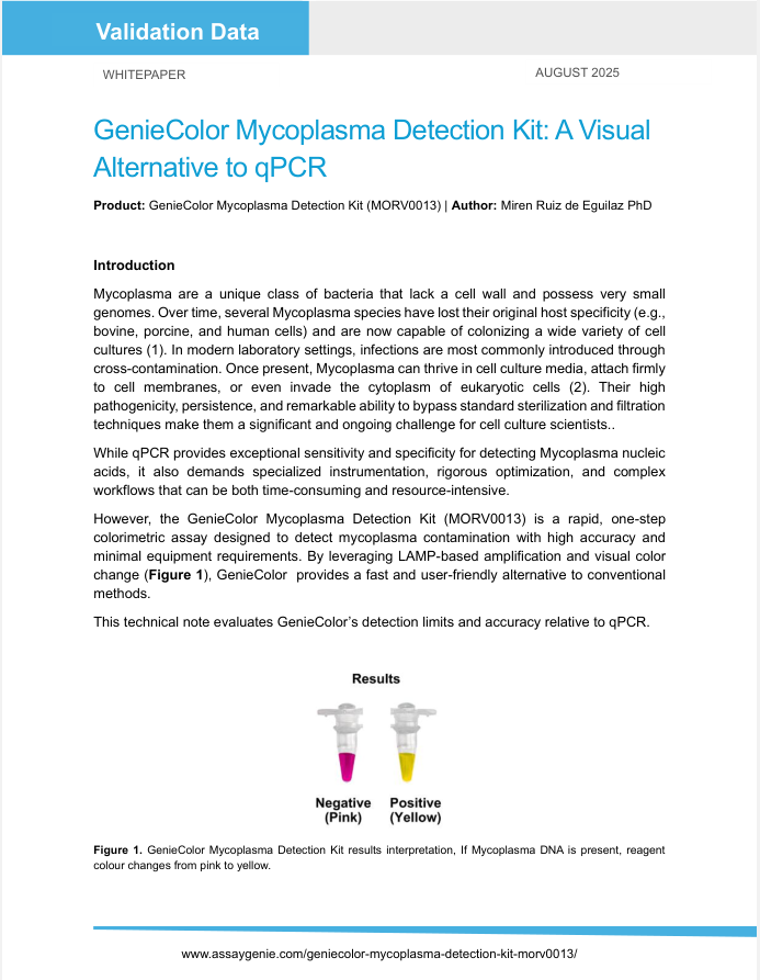
GenieColor is a mycoplasma detection kit for cell culture that gives a yes/no answer in one hour. Add 1 μl of culture supernatant to the pre-mixed LAMP reagent, incubate at 65°C in a water bath or PCR instrument, and read the result by eye: pink is negative, yellow is positive. No qPCR instrument, gel or plate reader is needed.
The kit detects 40 mycoplasma species, including the eight contaminants behind most cell-line infections (M. hyorhinis, M. fermentans, M. arginini, M. orale, M. hominis, M. salivarium, M. pirum and A. laidlawii), down to 100 copies/μl. Positive and negative controls are included, and an anti-contamination system prevents false positives from amplification aerosols. It works with adherent and suspension cells, conditioned media and serum.
Available in 25 and 50 tests, with a 10-test sample pack. Cited in peer-reviewed research and validated across laboratory conditions in two Assay Genie technical notes (see Citations and Technical Notes below). For research use only.
| Visual color change | Identify mycoplasma contamination with a pink-to-yellow color change. | |
| 1-Step protocol | Incubate 1μl cell culture supernatant with the reaction system for 60 min at 65°C. | |
| No specialized equipment needed | Simple set-up with no need for advanced instruments. | |
| Detect up to 40 mycoplasma species | Broad detection capability including 8 strains commonly found in cell cultures. |
Specifications
| Method | Loop-mediated isothermal amplification (LAMP), colorimetric |
| Readout | Visual: pink (negative) to yellow (positive) |
| Time | 60 min at 65°C, single step |
| Sample | 1 μl culture supernatant (adherent or suspension cells, ≥72 h in culture, ~90% confluence); media and serum compatible |
| Species detected | 40 mycoplasma species, including the 8 common cell-culture contaminants |
| Limit of detection | 100 copies/μl (positive-control plasmid) |
| Controls | Positive (M. hyorhinis plasmid, 10 ng/μl) and negative, included |
| Equipment | Water bath or PCR instrument; no qPCR, gel or reader |
| Pack sizes | 25 or 50 tests (10-test sample pack) |
| Storage / shipping | −30 to −15°C / ≤0°C |
| Catalogue number | MORV0013 |
How GenieColor compares with other mycoplasma tests
| GenieColor (this kit) | MycoGenie Rapid (MORV0011) | PCR kits | Luminescence enzyme kits | DNA staining (Hoechst/DAPI) | |
| Readout | Colour change, by eye | Colour change, by eye | Gel band | Luminometer | Fluorescence microscope |
| Time to result | 1 h | 1 h | 2–3 h including gel | About 20 min | 1–3 days with indicator cells |
| Equipment | Water bath | Water bath | Thermal cycler and electrophoresis | Luminometer | Microscope and indicator cell line |
| Species-specific | Yes, 40 species, LOD 100 copies/μl | Yes, 28 species, LOD 500 cfu/μl | Yes | No (measures enzyme activity) | No |
| Best for | Routine screening of many cell lines; broadest species coverage | Labs standardised on MORV0011; cited in Nature 2024 and Nature Metabolism 2024 | Confirmatory testing with a documented gel result | Fast go/no-go where a luminometer is available | Labs already running indicator cultures |
| Pyziac, S, A. et al. | Optimising an Experimental In Vitro Blood-Brain Barrier Model for Cancer Migration Studies | Master thesis, Aalborg University, Denmark 2026 | View Citation |
| Ragni, E., et al. | Matrix Protective and Anti-Inflammatory In Vitro Potential of Adipose Mesenchymal Stromal Cell-Derived EVs Coupled With Platelet-Derived Fibrin Gel | Frontiers in Bioscience-Landmark 2026 | View Citation |
Technology
The GenieColor kit utilizes an advanced isothermal amplification technology to detect mycoplasma contamination in cell cultures with a pink-to-yellow visual color change. It offers several benefits including ease of use, clear results, high sensitivity, strong specificity, and anti-contamination features. By adding 1 μl of cell culture supernatant to the reaction system and incubating at 65°C for one hour, results can be visually observed. If mycoplasma contamination is present, the reaction solution changes from pink to yellow, providing a clear color contrast. Additionally, the reagent incorporates an anti-contamination system to prevent false positives from amplification product aerosols, ensuring accurate and reliable results. The GenieColor Mycoplasma Detection kit can identify 40 mycoplasma species, including 8 commonly found in cell cultures, and is compatible with a wide range of adherent cells, suspension cells, media, and serum.
Components
| COMPONENTS | 10 Runs | 25 Runs | 50 Runs |
| GenieColor LAMP Mix | 200 μl | 500 μl | 2 × 500 μl |
| Positive Control | 10 μl | 15 μl | 2 × 15 μl |
| Mineral Oil | 200 μl | 500 μl | 2 × 500 μl |

Storage
Store at -30°-15°C and transport at ≤0°C.
-
Collect the cell culture supernatant
- Adherent cells: directly collect the supernatant. Cells should remain in culture for at least 72 h undisturbed prior to screening and reach 90% confluence. After 72 h, mycoplasma content in supernatant is relatively high and can be detected easily.
- Suspension cells: collect the supernatant after centrifugation at 2,300 rpm (500 × g) for 5 min. Cells should remain in culture for at least 72 h undisturbed prior to screening. After 72 h, mycoplasma content in supernatant is relatively high and can be detected easily.
-
Preparation of reaction system
- Thaw the GenieColor LAMP Mix from -30°-15°C and mix thoroughly.
- Prepare the reaction system according to the following table:
| Components | Test Sample | Negative Control* | Positive Control |
| GenieColor LAMP Mix | 19 μl | 19 μl | 19 μl |
| Test Sample | 1 μl | - | - |
| Nuclease-free ddH2O | - | 1 μl | - |
| Positive Control | - | - | 1 μl |
| Total | 20 μl | 20 μl | 20 μl |
- Vortex and briefly centrifuge to collect at the bottom of the tube, ensuring no bubbles in the reaction system. Add 20 μl of mineral oil if using a water bath for incubation.
-
Reaction
- Incubate at 65°C for 60 min in a PCR instrument or water bath (make sure water bath is calibrated prior to use).
-
Results
- After the reaction is completed, observe the color in a bright environment. Pink represents negative and yellow represents positive.
- Do not open the lid of detection tubes to prevent false positives due to aerosol contamination.
Q1: Do I need a PCR machine to run GenieColor?
- No. The reaction is isothermal, so a calibrated water bath at 65°C is enough; a PCR instrument works too. You read the result by eye, so no gel, plate reader or luminometer is needed.
Q2: How does GenieColor differ from a PCR mycoplasma kit?
- PCR kits amplify by thermal cycling and read the result on an agarose gel, which typically takes 2–3 hours including electrophoresis. GenieColor uses LAMP amplification with a colour indicator, so the result appears in the tube after 60 minutes with no gel, and the reagent is more tolerant of culture-medium inhibitors than PCR. Assay Genie also offers the MycoGenie Rapid Mycoplasma Detection Kit (MORV0011), the same 1-hour visual format covering 28 species, which is cited in Nature 2024 and Nature Metabolism 2024.
Q3: What is the sensitivity of the kit (minimum detection limit)?
- The kit can detect mycoplasma positive plasmid at a minimum concentration of 100 copies/μl.
Q4: Which mycoplasma species does it detect?
- The kit detects 40 mycoplasma species, including the eight most common cell-culture contaminants: M. hyorhinis, M. fermentans, M. arginini, M. orale, M. hominis, M. salivarium, M. pirum and A. laidlawii. The full list is in the last question below.
Q5: What sample do I use, and how should I prepare it?
- Use 1 μl of culture supernatant. Leave cells undisturbed in culture for at least 72 h and, for adherent lines, reach about 90% confluence so that mycoplasma in the supernatant is high enough to detect. For suspension cultures, centrifuge at 500 × g for 5 min and use the supernatant.
Q6: What does it mean if the reaction liquid color is between pink and yellow?
- A color between pink and yellow indicates a weak positive. It is recommended to repeat the test. If the repeated test is positive or weak positive, it is considered positive. If the repeated test is negative, it is considered negative.
Q7: How is accuracy ensured?
- The product includes both negative and positive controls to ensure accurate results. It has been tested against 40 different mycoplasma species, including 8 common mycoplasma contaminants.
Q8: What is the detection principle of this kit?
- The kit utilizes loop-mediated isothermal amplification (LAMP) technology. The reagent is pre-mixed with Bst isothermal amplification enzyme, mycoplasma-specific primers (inner and outer primers), and an optimized buffer. After adding the template, the LAMP reaction begins. If mycoplasma DNA is present, the reagent color changes from pink to yellow. A negative result remains pink, while a positive result turns yellow.
Q9: What sequence is used in the positive control?
- The positive control contains a plasmid with the M. hyorhinis sequence at a concentration of 10 ng/μl.
Q10: Does the positive control of the kit cause contamination?
- No, the positive control is a mycogenic plasmid, which does not cause contamination.
Q11: Why does the reaction solution change color after adding the supernatant of the medium?
- The medium might interfere with the color change. If it does, then use the following treatment method:
| Treatment Method: |
|
Q12: What 40 Mycoplasma species are detected using this kit?
| Number | Mycoplasma Name | Number | Mycoplasma Name | Number | Mycoplasma Name | Number | Mycoplasma Name |
| 1 | M. hyorhinis* | 11 | M. gallisepticum | 21 | M. testudineum | 31 | M. genitalium |
| 2 | M. fermentans* | 12 | M. synoviae | 22 | M. columbinum | 32 | M. iguanae |
| 3 | M. arginini* | 13 | M. auris | 23 | M. cricetuli | 33 | M. leopharyngis |
| 4 | M. orale* | 14 | M. crocodyli | 24 | M. gallinarum | 34 | M. lipofaciens |
| 5 | M. hominis* | 15 | M. iners | 25 | M. opalescens | 35 | M. hyosynoviae |
| 6 | M. salivarium* | 16 | M. bovigenitalium | 26 | M. moatsii | 36 | M. falconis |
| 7 | M. pirum* | 17 | M. simbae | 27 | M. alkalescens | 37 | M. meleagridis |
| 8 | A. laidlawii* | 18 | M. arthritidis | 28 | M. sphenisci | 38 | M. mobile |
| 9 | M. pneumoniae | 19 | M. alvi | 29 | M. agalactiae | 39 | M. cloacale |
| 10 | M. bovis | 20 | M. primatum | 30 | M. cynos | 40 | M. spermatophilum |