Human VIP ELISA kits are used to detect Vasoactive Intestinal Peptide (VIP) in serum, plasma, tissue homogenates, cell lysates, cell culture supernatants and other biological samples. VIP is a neuropeptide that acts through the PACAP subfamily of class B1 G protein-coupled receptors, VPAC1 and VPAC2. It is abundantly expressed throughout the central and peripheral nervous systems, including the gut, where it exerts neuroprotective and immune-modulatory roles. VIP causes vasodilation, lowers arterial blood pressure, stimulates myocardial contractility, increases glycogenolysis and relaxes smooth muscle of the trachea, stomach and gallbladder. It is processed from a precursor that also yields the peptides PHM-27 and PHV-42, which are also vasodilatory, and PHM-27 is a potent agonist of the calcitonin receptor. Elevated VIP levels are characteristic of VIP-secreting tumours causing watery diarrhoea, and VIP is studied in circadian biology, inflammation and neuroendocrine research.
Serum, Plasma, Tissue Homogenates, Cell Lysates, Cell Culture Supernates And Other Biological Fluids
Kit Components:
Component
Quantity
Storage
48T
96T
Pre-Coated Microplate
6 strips x 8 wells
12 strips x 8 wells
4°C/-20°C
Standard (Lyophilized)
1 vial
2 vials
4°C/-20°C
Biotinylated Antibody (100×)
60 μL
120 μL
4°C/-20°C
Streptavidin-HRP (100×)
60 μL
120 μL
4°C/-20°C
Standard / Sample Diluent Buffer
10 mL
20 mL
4°C/-20°C
Biotinylated Antibody Diluent
6 mL
12 mL
4°C/-20°C
HRP Diluent
6 mL
12 mL
4°C/-20°C
Wash Buffer (25×)
10 mL
20 mL
4°C/-20°C
TMB Substrate Solution
6 mL
10 mL
4°C/-20°C (store in dark)
Stop Reagent
3 mL
6 mL
4°C/-20°C
Plate Covers
1 piece
2 pieces
RT
The test principle applied in this kit is Sandwich enzyme immunoassay. The microtiter plate provided in this kit has been pre-coated with an antibody specific to Human VIP. Standards or samples are added to the appropriate microtiter plate wells then with a biotin-conjugated antibody specific to Human VIP. Next, Avidin conjugated to Horseradish Peroxidase (HRP) is added to each microplate well and incubated. After TMB substrate solution is added, only those wells that contain Human VIP, biotin-conjugated antibody and enzyme-conjugated Avidin will exhibit a change in color. The enzyme-substrate reaction is terminated by the addition of sulphuric acid solution and the color change is measured spectrophotometrically at a wavelength of 450nm ± 10nm. The concentration of Human VIP in the samples is then determined by comparing the OD of the samples to the standard curve.
Standard Curve:
Concentration (pg/mL)
OD
Corrected OD
500.00
2.096
2.003
250.00
1.603
1.510
125.00
1.175
1.082
62.50
0.817
0.724
31.25
0.585
0.492
15.63
0.313
0.220
7.82
0.252
0.159
0.00
0.093
0.000
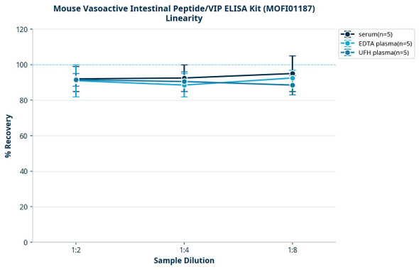
Linearity:
Matrix
1:2
1:4
1:8
1:16
Serum (n=5)
85-105%
87-98%
91-102%
86-102%
EDTA Plasma (n=5)
86-94%
93-102%
87-96%
92-105%
Heparin Plasma (n=5)
80-101%
87-96%
96-105%
95-103%
Recovery:
Matrix
Recovery range
Average
Serum (n=5)
90-107%
98%
EDTA Plasma (n=5)
87-99%
93%
Heparin Plasma (n=5)
80-95%
87%
Precision:
Intra-assay Precision (Precision within an assay)
Intra-assay Precision (Precision within an assay):CV%<8%
Three samples of known concentration were tested twenty times on one plate to assess intra-assay precision.
Inter-assay Precision (Precision between assays)
Inter-assay Precision (Precision between assays):CV%<10%
Three samples of known concentration were tested in forty separate assays to assess inter-assay precision.
*Note: The below protocol is a sample protocol. Protocols are specific to each batch/lot. For the correct instructions please follow the protocol included in your kit.
Step
Protocol
1.
After the kit is equilibrated at room temperature, add 100 µL of Standard Working Buffer (gradually diluted according to the instructions) or 100 µL of sample to each well, and incubate at 37°C for 80 minutes.
2.
Discard the liquid in the plate, add 200 µL 1× Wash Buffer to each well, and wash the plate 3 times. After pat it dry against clean absorbent paper, add 100 µL Biotinylated Antibody Working Solution (1×) to each well, incubate at 37°C for 50 minutes.
3.
Discard the liquid in the plate, add 200 µL 1× Wash Buffer to each well, and wash the plate 3 times. After pat it dry against clean absorbent paper, add 100 µL 1× Streptavidin-HRP Working Solution to each well, incubate at 37°C for 50 minutes.
4.
Discard the liquid in the plate, add 200 µL 1× Wash Buffer to each well, and wash the plate 5 times. After pat it dry against clean absorbent paper, add 90 µL TMB Substrate Solution to each well, incubate at 37°C for 20 minutes in the dark.
5.
Add 50 µL Stop Solution to each well, shake plate on a plate shaker for 1 minute to mix. Record the OD at 450 nm immediately, calculation of the results.
When carrying out an ELISA assay it is important to prepare your samples in order to achieve the best possible results. Below we have a list of procedures for the preparation of samples for different sample types.
Sample Type
Protocol
Serum
Samples should be collected into a serum separator tube. After clotting for 2 hours at room temperature or overnight at 4°C, and then centrifuging at 1000 × g for 20 minutes. Assay freshly prepared serum immediately or store samples in aliquot at -20°C or -80°C for later use. Avoid repeated freeze-thaw cycles.
Plasma
Collect plasma using EDTA or heparin as an anticoagulant. Centrifuge samples at 1000 × g and 2-8°C for 15 minutes within 30 minutes of collection. Remove plasma and assay immediately or store samples in aliquot at -20°C or -80°C for later use. Avoid repeated freeze-thaw cycles.
Tissue homogenates
1. Rinse the tissues in pre-cooled PBS to completely remove excess blood, and weigh them before homogenization. 2. Mince the tissues and homogenize in fresh lysis buffer (PBS for most tissues). Use a glass homogenizer on ice. 3. Ultrasound the suspension until the solution is clear. 4. Centrifuge for 5 minutes at 10000 × g, collect the supernatant and assay immediately or store at ≤ -20°C.
Cell lysates
1. Wash adherent cells with PBS, detach with trypsin, and centrifuge at 1000 × g for 5 minutes. 2. Wash cells 3 times in PBS. 3. Resuspend cells in fresh lysis buffer at 107 cells/mL. Ultrasound if necessary. 4. Centrifuge at 1500 × g for 10 minutes at 2-8°C to remove debris. Assay immediately or store at ≤ -20°C.
Urine
Collect mid-stream first urine of the day directly into a sterile container. Centrifuge to remove particulate matter. Assay immediately or aliquot and store at ≤ -20°C. Avoid repeated freeze-thaw cycles.
Saliva
Collect saliva using a collection device. Centrifuge at 1000 × g for 15 minutes at 2-8°C. Remove particulates and assay immediately or aliquot and store at ≤ -20°C. Avoid repeated freeze-thaw cycles.
Feces
Dry feces weighing more than 50 mg were collected. Wash with PBS (w:v = 1:9). Sonicate and centrifuge at 5000 × g for 10 minutes. Collect the supernatant and assay immediately.
CSF (Cerebrospinal fluid)
Remove particulates by centrifugation. Assay immediately or aliquot and store at ≤ -20°C. Avoid repeated freeze-thaw cycles.
Cell culture supernatant
Centrifuge samples at 1000 × g for 20 minutes. Collect the supernatant and assay immediately or store at -20°C or -80°C. Avoid repeated freeze-thaw cycles.