Description
α-Glucosidase (α-GC) Activity Assay Kit
The α-Glucosidase (α-GC) Activity Assay Kit (MAES0460) measures α-GC enzyme activity in tissue and fungal homogenates using a colorimetric readout at 400 nm. The assay is designed for research use in carbohydrate metabolism and glycogen storage studies. Activity is calculated in U/g protein, so results can be normalized to sample protein concentration.
α-Glucosidase (α-GC), also known as α-D-glucoside hydrolase, is widely distributed in nature and is found in almost all organisms, with a wide variety of species and differing properties. It performs important physiological functions in carbohydrate metabolism in animals, plants, and microorganisms. Its activity is needed for normal glycogen metabolism, and reduced activity disrupts that process.
Measuring α-GC activity supports research in carbohydrate and glycogen metabolism. Deficiency of α-GC leads to severe glycogen metabolism disorders and excessive glycogen accumulation, which can cause Pompe disease (an autosomal recessive inherited glycogen storage disease, also known as glycogenosis type II). In human tissues, long-term low α-GC activity can cause muscle fiber destruction and muscle atrophy. The kit is categorized under glycolysis and lipid metabolism research.
The assay is a colorimetric method. The substrate reaction catalyzed by α-glucosidase generates a chromogenic substance with maximum absorption at 400 nm, so enzyme activity is reflected by the optical density (OD) at 400 nm. Each well uses a working solution prepared from 8 µL of substrate and 112 µL of buffer solution. Standard wells receive 20 µL of standard plus 120 µL of buffer solution, and sample wells receive 20 µL of sample plus 120 µL of working solution. After mixing for 5 s, the OD is read (A1), the plate is incubated at 37°C for 30 min, and the OD is read again (A2). The calculation uses ΔA400 = A2 − A1.
The kit is intended for tissue and fungus samples. The manual lists dilution factors for 10% homogenates of mouse small intestine, rat kidney, and mouse kidney tissue (1 to 2), and for orange, pear, and apple seeds, apricot abalone mushroom, and corn tissue (1). Tissues are washed in cold PBS (0.01 M, pH 7.4), homogenized at 4°C, and centrifuged at 12000×g for 15 min at 4°C to remove insoluble material. The supernatant is kept on ice until detection. Reagents are stored at -20°C for 12 months, with the substrate and standard protected from light. The working solution should be used within 1 day.
Performance was characterized with rat kidney samples. Intra-assay CV ranged from 4.4% to 5.7%, and inter-assay CV ranged from 4.2% to 6.6%. Recovery at low, middle, and high standard concentrations was 98%, 106%, and 110%, with an average of 105%. The analytical sensitivity is 0.35 U/L, and the detection range is 0.35-33.43 U/L. The assay time is 50 min.
| Product name: | α-Glucosidase (α-GC) Activity Assay Kit |
| SKU: | MAES0460 |
| Sizes: | 48T (32 samples) / 96T (80 samples) |
| Sample type: | Tissue, fungal samples |
| Detection method: | Colorimetric |
| Instrument: | Microplate reader (390-405 nm; optimal 400 nm) |
| Assay time: | 30 min at 37°C |
| Research area: | Enzymes, Carbohydrate Metabolism |
| Intended use: | Research Use Only |
| Sensitivity: | 0.35 U/L |
| Detection range: | 0.35-33.43 U/L |
| Standard curve range: | 0-1 mmol/L (standard solution 10 mmol/L) |
| Intra-assay CV: | 4.4-5.7% |
| Inter-assay CV: | 4.2-6.6% |
| Recovery rate: | 105% (average) |
| Sample volume: | 20 µL per well (tissue/fungal homogenate) |
| Working solution volume: | 120 µL per well |
| Activity unit: | 1 U = 1 µmol product per min at 37°C per g sample protein |
- Colorimetric detection: chromogenic product read at 400 nm (microplate reader, 390-405 nm)
- Detection range 0.35-33.43 U/L; analytical sensitivity 0.35 U/L
- Validated for tissue and fungal samples; 20 µL sample per well
- Assay time: 30 min incubation at 37°C after initial read
- Intra-assay CV 4.4-5.7% and inter-assay CV 4.2-6.6%
- Supplied as 48T (32 samples) or 96T (80 samples)
The substrate is hydrolyzed by α-glucosidase to generate a chromogenic product with maximum absorption at 400 nm. α-GC activity is determined by measuring the change in optical density at 400 nm (ΔA400 = A2 − A1) over a 30 min incubation at 37°C, and is calculated against a blank-corrected standard curve.
- Measuring α-GC activity in mammalian tissue homogenates for carbohydrate metabolism studies
- Investigating α-GC activity in fungal samples
- Comparing α-GC activity across tissues, species, or experimental conditions, normalized to protein concentration
- Supporting research on glycogen storage and glycogen metabolism disorders such as Pompe disease models
| Component | Quantity | Storage |
| Extraction Solution (Reagent 1) | 48T: 50 mL × 1 vial; 96T: 50 mL × 2 vials | -20°C, 12 months |
| Buffer Solution (Reagent 2) | 48T: 7 mL × 1 vial; 96T: 14 mL × 1 vial | -20°C, 12 months |
| Substrate (Reagent 3) | 48T: 0.5 mL × 1 vial; 96T: 1 mL × 1 vial | -20°C, 12 months, protect from light |
| Standard Solution, 10 mmol/L (Reagent 4) | 48T: 1 mL × 1 vial; 96T: 1 mL × 2 vials | -20°C, 12 months, protect from light |
| Microplate | 48T: 48 wells; 96T: 96 wells | No requirement |
| Plate sealer | 2 pieces | No requirement |
| Sample layout sheet | 1 piece | No requirement |
- Microplate reader capable of 390-405 nm reading (optimal 400 nm)
- 37°C incubator
- Dounce homogenizer
- Refrigerated centrifuge capable of 12000 × g
- Ice and 4°C cold room or refrigerator
- PBS (0.01 M, pH 7.4) for tissue washing
- Double-distilled water for standard dilutions
- Protein concentration assay for sample supernatant (for U/g protein normalization)
- Precision pipettes and tips
Tissue and fungus samples
- Harvest the amount of tissue or fungus needed for each assay (initial recommendation: 20 mg).
- Wash the tissue or fungus in cold PBS (0.01 M, pH 7.4).
- Homogenize 20 mg of tissue or fungus in 180 µL of extraction solution with a Dounce homogenizer at 4°C to yield a 10% homogenate.
- Centrifuge at 12000×g for 15 min at 4°C to remove insoluble material.
- Collect the supernatant and keep it on ice for detection.
- Determine the protein concentration of the supernatant with a separate protein quantification assay, as it is required for the calculation.
- Dilution: the recommended dilution factors (for reference only) are 1 to 2 for 10% mouse small intestine tissue homogenate, 10% rat kidney tissue homogenate, and 10% mouse kidney tissue homogenate; and 1 for 10% orange seed, 10% apricot abalone mushroom tissue, 10% pear seed, 10% corn tissue, and 10% apple seed homogenates.
- Dilute samples with extraction solution. For other sample types, perform a pretest to confirm the dilution factor.
Reagent preparation
- Equilibrate all reagents to room temperature before use.
- Working solution: per well, mix 8 µL substrate with 112 µL buffer solution (120 µL total). Use within 1 day.
- 1 mmol/L standard solution: mix 100 µL of 10 mmol/L standard solution with 900 µL double-distilled water. Prepare fresh before testing and protect from light.
- Standard curve: prepare a fresh set of standards by diluting 1 mmol/L standard with double-distilled water to 0, 0.1, 0.2, 0.3, 0.4, 0.5, 0.7, and 1 mmol/L (for example, 0, 20, 40, 60, 80, 100, 140, and 200 µL of 1 mmol/L standard, brought to 200 µL with water). Discard working dilutions after use.
Sample preparation (tissue and fungal)
- Harvest the tissue or fungal amount needed for each assay (initial recommendation: 20 mg).
- Wash tissue or fungus in cold PBS (0.01 M, pH 7.4).
- Homogenize 20 mg tissue or fungus in 180 µL extraction solution with a Dounce homogenizer at 4°C.
- Centrifuge at 12000 × g for 15 min at 4°C to remove insoluble material. Collect the supernatant and keep on ice.
- Determine the protein concentration of the supernatant using a separate protein assay.
- Dilute samples as needed using extraction solution. Recommended dilution factors: 1-2 for 10% mouse small intestine, rat kidney, and mouse kidney homogenates; 1 for 10% orange seed, apricot abalone mushroom, pear seed, corn, and apple seed homogenates. For other sample types, perform a pretest to confirm the dilution factor.
Assay procedure
- Add 20 µL of standard to standard wells and 20 µL of diluted sample to sample wells.
- Add 120 µL buffer solution to standard wells. Add 120 µL working solution to sample wells.
- Mix fully on the microplate reader for 5 s and read the OD at 400 nm (A1).
- Incubate at 37°C for 30 min, then read the OD at 400 nm (A2) for each well.
α-GC activity (U/gprot) = (ΔA400 - b) ÷ a ÷ T × f ÷ Cpr × 1000*
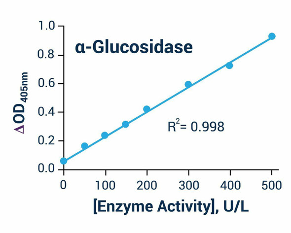
- a and b are the slope and intercept of the standard curve y = ax + b, where y is the absolute OD (blank-subtracted mean OD of each standard) and x is the standard concentration in mmol/L.
- ΔA400 = A2 − A1, where A1 is the OD at 400 nm before incubation and A2 is the OD at 400 nm after 30 min at 37°C.
- T is the reaction time, 30 min.
- f is the dilution factor of the sample before testing. Multiply by the dilution factor; use 1 for undiluted samples.
- Cpr is the protein concentration of the sample in gprot/L.
- 1000 converts 1 mmol/L to 1000 µmol/L.
- Units: 1 unit is defined as the amount of enzyme in 1 g of sample protein that hydrolyzes substrate to produce 1 µmol of product in 1 min at 37°C. Results are reported in U/gprot.
- Example from the manual: a 10% mouse small intestine homogenate diluted 2-fold, with standard curve y = 0.4766x − 0.0001, ΔA400 = 0.312, and Cpr = 9.06 gprot/L, gives (0.312 + 0.0001) ÷ 0.4766 ÷ 30 ÷ 9.06 × 2 × 1000 = 4.82 U/gprot.
What sample types can be tested with this kit?
The kit detects α-GC activity in tissue and fungus samples. For other sample types, perform a pretest to confirm the dilution factor.
How should the kit components and samples be stored?
Reagents are stored at -20°C for 12 months, with the substrate and standard protected from light. The working solution must be used within 1 day, and the homogenate supernatant should be kept on ice until detection.
What should I do if my sample is outside the detection range?
The detection range is 0.35-33.43 U/L. If the concentration is not within this range, take an extra dilution or concentration of the sample and reassay it.
What factors can affect results?
Results are closely related to reagent condition, operation, and environment. Reagents from different kits must not be mixed, and a pretest is recommended for samples not listed in the manual.
What instrument settings are required?
Use a microplate reader set to 390-405 nm, with 400 nm as the optimum wavelength. The incubation step is performed at 37°C.
How long does the assay take?
The assay time is 50 min, including a 30-min incubation at 37°C after the initial OD reading.
| Storage: | -20°C; protect light-sensitive components from light |
| Shelf Life: | 12 months |