Description
GSH/GSSG Colorimetric Assay Kit
The GSH/GSSG Colorimetric Assay Kit (MAES0086) quantifies total glutathione (T-GSH, GSH plus GSSG) and oxidized glutathione (GSSG) in serum, plasma, animal tissue, red blood cells, and cultured cells. Total glutathione is measured by a glutathione reductase-coupled DTNB reaction read at 412 nm on a microplate reader. GSSG is determined after GSH is removed from the sample with a GSH scavenger, and GSH is calculated by difference. Researchers use the kit to assess cellular redox status in oxidative stress studies.
Glutathione occurs in two forms in biological samples: reduced glutathione (GSH) and oxidized glutathione (GSSG). This kit measures total glutathione (T-GSH, defined as GSSG plus GSH) and GSSG directly. Reduced GSH is not measured separately but is derived by calculation as T-GSH minus twice the GSSG content. Both analytes are reported in the kit's calculation section, which allows the reduced and oxidized fractions to be compared within one sample set.
The product page lists oxidative stress, cuproptosis, and ferroptosis as research areas for this kit. Appendix III cites a 2024 study of selenocystine protection against cadmium-induced liver damage and a study of crosstalk between ferroptosis and necroptosis in cerebral ischemia/reperfusion injury, both of which measure glutathione-related endpoints. These citations place the kit in liver injury and cerebral injury research, as well as in studies of redox-dependent cell death.
The assay is colorimetric. GSSG is reduced to GSH by glutathione reductase, and GSH reacts with DTNB to produce GSSG and yellow TNB, so the amount of yellow TNB reflects total glutathione. Absorbance is read at 412 nm on a microplate reader set to 405-415 nm. For GSSG, GSH is first removed from the sample with a GSH scavenger, and the same reaction is then applied. In the operating steps, 10 µL of standard or sample is mixed with 150 µL reaction working solution and incubated for 5 min, then 50 µL substrate working solution is added and the plate is incubated for 25 min before reading.
The kit is validated for serum, plasma, animal tissue, whole blood, red blood cells, and cultured cells. Serum and plasma are deproteinized with protein precipitator at a 1:4 sample-to-precipitator ratio, and tissue and cell lysates are prepared in precipitator at 4°C. Supernatants are kept on ice until assayed. Enzyme stock solution must be kept on ice during use, while other reagents are equilibrated to room temperature. Working solutions are stable for 24 h at 2-8°C, so they should be prepared fresh on the day of the assay.
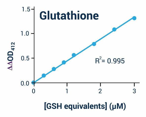
The analytical sensitivity is 0.36 µmol/L, determined by adding two standard deviations to the mean OD of the zero standard assayed 20 times. The detection range is 0.36-30 µmol/L. Intra-assay precision is 0.6% CV and inter-assay precision is 3.9% CV on the product page. Spike recovery averaged 97% across three concentrations, with individual recoveries of 94-99%. Standard curve values in the manual are provided for reference only and vary with operator, pipetting, and temperature.
| Product Name: | GSH/GSSG Colorimetric Assay Kit |
| SKU: | MAES0086 |
| Size: | 96 Assays |
| Analytes: | T-GSH (total glutathione), GSSG; GSH by calculation |
| Sample Types: | Serum, plasma, animal tissue, red blood cells, cultured cells |
| Detection Method: | Colorimetric (DTNB-based) |
| Instrument: | Microplate reader (405-415 nm; optimum 412 nm) |
| Assay Time: | 100 min (1 h 40 min) |
| Research Area: | Oxidative stress; cuproptosis; ferroptosis |
| Research Use Only: | Yes |
| Sensitivity: | 0.36 µmol/L T-GSH |
| Detection Range: | 0.36-30 µmol/L T-GSH |
| Recovery Rate: | 97% (average) |
| Intra-assay CV: | 0.6% |
| Inter-assay CV: | 3.9% |
| Sample Volume: | 100 µL |
| Assay Time: | 100 min (1 h 40 min) |
- Colorimetric detection at 412 nm on a microplate reader (405-415 nm)
- Measures T-GSH and GSSG; reduced GSH calculated from the two values
- Sensitivity 0.36 µmol/L; detection range 0.36-30 µmol/L T-GSH
- Intra-assay CV 0.6% and inter-assay CV 3.9%; average recovery 97%
- Assay time 100 min (1 h 40 min)
- Supplied as 96 Assays
GSSG is reduced to GSH by glutathione reductase, and GSH reacts with DTNB to produce GSSG and yellow TNB. The amount of total glutathione (GSSG+GSH) determines the amount of yellow TNB, so total glutathione is calculated from the OD at 412 nm. GSSG is determined by first removing GSH from the sample with a GSH scavenger and then applying the same reaction principle.
- Measurement of total glutathione (T-GSH) and GSSG to assess cellular redox status
- Oxidative stress studies in serum, plasma, animal tissue, and cultured cells
- Calculation of reduced GSH content from T-GSH and GSSG values
- Studies of apoptosis-related signal transduction and cell homeostasis
- Research on cuproptosis and ferroptosis
| Component | Quantity | Storage |
| Reagent 1: Buffer Solution | 45 mL × 1 vial (96 Assays) | -20°C, 12 months |
| Reagent 2: Standard | 6.13 mg × 1 vial (96 Assays) | -20°C, 12 months |
| Reagent 3: Protein Precipitator | 50 mL × 2 vials (96 Assays) | -20°C, 12 months |
| Reagent 4: Enzyme Stock Solution | 80 µL × 1 vial (96 Assays) | -20°C, 12 months |
| Reagent 5: Chromogenic Agent | 1 mL × 1 vial (96 Assays) | -20°C, 12 months, protect from light |
| Reagent 6: GSH Scavenger Auxiliary Solution | 1.1 mL × 1 vial (96 Assays) | -20°C, 12 months |
| Reagent 7: GSH Scavenger | 0.1 mL × 1 vial (96 Assays) | -20°C, 12 months, protect from light |
| Reagent 8: Substrate Powder | 1 vial (96 Assays) | -20°C, 12 months, protect from light |
| Microplate | 96 wells × 1 (96 Assays) | No special requirement |
| Plate Sealer | 2 pieces (96 Assays) | No special requirement |
| Sample Layout Sheet | 1 piece (96 Assays) | No special requirement |
- Microplate reader (405-415 nm; optimum 412 nm)
- Micropipettor
- Centrifuge (including a 0.5 mL EP tube-compatible rotor for GSSG pretreatment)
- Incubator (room temperature or 25°C)
- Vortex mixer
- Ice bucket
- Double-distilled water
- PBS (0.01 mol/L, pH 7.4)
- Absolute ethanol
- Dounce homogenizer or ultrasonic cell disruptor (for tissue or cell samples)
- Fume hood (for handling GSH scavenger)
Serum/plasma
- Prepare serum or plasma by the common method.
- Take 100 µL of sample and add 400 µL of protein precipitator.
- Mix fully by vortexing for 30 s, then stand for 5 min at 4°C.
- Centrifuge at 3100×g for 10 min.
- Collect the supernatant and keep it on ice for detection.
Whole blood
- Collect blood using heparin or EDTA as the anticoagulant.
- Take 100 µL of whole blood and add 400 µL of protein precipitator.
- Mix fully by vortexing for 30 s, then stand for 5 min at 4°C.
- Centrifuge at 3100×g for 10 min.
- Collect the supernatant and keep it on ice for detection.
Red blood cells
- Collect blood using heparin or EDTA as the anticoagulant.
- Centrifuge at 2000 rpm for 10 min immediately.
- Remove the plasma and leukocytic layer (upper layer) carefully.
- Take 100 µL of red blood cells and add 400 µL of protein precipitator.
- Mix fully by vortexing for 30 s, then stand for 5 min at 4°C.
- Centrifuge at 3100×g for 10 min.
- Collect the supernatant and keep it on ice for detection.
Tissue
- Harvest the amount of tissue needed for each assay (initial recommendation 20 mg).
- Wash the tissue in cold PBS (0.01 M, pH 7.4).
- Homogenize 20 mg of tissue in 180 µL of protein precipitator with a Dounce homogenizer at 4°C.
- Centrifuge at 10000×g for 10 min at 4°C to remove insoluble material.
- Collect the supernatant and keep it on ice for detection.
Cells (adherent or suspension)
- Harvest the number of cells needed for each assay (initial recommendation 1×10^6 cells).
- Wash cells with PBS (0.01 M, pH 7.4).
- Homogenize 1×10^6 cells in 200 µL of protein precipitator with an ultrasonic cell disruptor at 4°C.
- Centrifuge at 10000×g for 10 min at 4°C to remove insoluble material.
- Collect the supernatant and keep it on ice for detection.
Sample dilution
- Dilute samples with protein precipitator, which is the diluent specified in the manual.
- Recommended dilution factors (reference only): human plasma 1; 10% rat liver tissue homogenate 10-20; 10% rat kidney tissue homogenate 1; 10% rat heart tissue homogenate 10-20; 10% mouse brain tissue homogenate 2-5; HepG2 cells 1.
- For sample types not listed, run a pretest to confirm the dilution factor.
Sample preparation (serum or plasma)
- Mix 100 µL sample with 400 µL protein precipitator by vortexing for 30 s; stand 5 min at 4°C.
- Centrifuge at 3100×g for 10 min.
- Collect the supernatant and keep on ice for detection.
Sample preparation (tissue or cells)
- Homogenize 20 mg tissue in 180 µL protein precipitator at 4°C (initial recommendation), or 1×10^6 cells in 200 µL protein precipitator using an ultrasonic disruptor at 4°C.
- Centrifuge at 10000×g for 10 min at 4°C.
- Collect the supernatant and keep on ice for detection.
Standard curve preparation
- Dissolve one standard vial in 10 mL double-distilled water to make a 1 mmol/L stock.
- Dilute 20 µL of 1 mmol/L stock with 980 µL protein precipitator to make 20 µmol/L standard.
- Dilute the 20 µmol/L standard with protein precipitator to 0, 0.5, 1, 2, 5, 8, 10, and 15 µmol/L (for example, 0 µL: 400 µL precipitator for 0 µmol/L; 300 µL standard with 100 µL precipitator for 15 µmol/L).
- Prepare standards fresh and discard working dilutions after use.
Working solutions
- Reaction working solution: mix 1 µL enzyme stock solution, 20 µL chromogenic agent, and 519 µL buffer solution (540 µL total; 150 µL per well). Stable 24 h at 2-8°C.
- Substrate working solution: mix 5 µL substrate stock (substrate powder dissolved in 100 µL double-distilled water) with 395 µL buffer solution (400 µL total; 50 µL per well).
- GSH scavenger auxiliary working solution: dilute 20 µL GSH scavenger auxiliary solution with 20 µL double-distilled water.
- GSH scavenger working solution: dilute 5 µL GSH scavenger with 45 µL absolute ethanol.
T-GSH measurement
- Add 10 µL of each standard or sample to the corresponding wells.
- Add 150 µL reaction working solution to each well; incubate 5 min at room temperature or 25°C.
- Add 50 µL substrate working solution to each well and mix for 5 s on the microplate reader.
- Incubate 25 min at room temperature or 25°C; read OD at 412 nm.
Serum/plasma/whole blood/red blood cells: T-GSH content (µmol/L) = (ΔA1 − b1) ÷ a1 × 2* × 5** × f; GSSG content (µmol/L) = (ΔA2 − b2) ÷ a2 × 5** × f; Reduced GSH content = T-GSH content − 2 × GSSG content
- Standard curves are plotted as absolute OD (y) against standard concentration in µmol/L (x), giving y = ax + b. Absolute OD is the mean standard OD minus the mean blank OD (Standard #1).
- a1 and b1 are the slope and intercept of the T-GSH standard curve; a2 and b2 are the slope and intercept of the GSSG standard curve.
- ΔA1 is the sample OD412 minus the sample blank OD412 for T-GSH; ΔA2 is the sample OD412 minus the sample blank OD412 for GSSG.
- 2* applies only to T-GSH: GSSG is the standard, so the result is multiplied by 2 to convert to GSH. No factor of 2 is applied to the GSSG result.
- 5** is the dilution multiple of the sample in the sample preparation step (100 µL sample plus 400 µL protein precipitator gives 5).
- f is the additional dilution factor applied to the sample before testing; use 1 if the sample was not diluted further.
- Tissue results are expressed per gram of fresh weight (µmol/g). The manual divides by the fresh weight (m) and the precipitator volume (V1) used in tissue preparation, as shown in the tissue section of the calculation.
- Cell results are expressed per 1×10^6 cells (µmol/10^9 in the manual's notation, which uses the cell number 1×10^6). The manual divides by the precipitator volume (V2) used in cell preparation.
- Example from the manual: 10% rat liver homogenate diluted 20-fold with protein precipitator, 10 µL assayed, gave T-GSH of 4.36 µmol/g and GSSG of 0.52 µmol/g.
What sample types can be tested with this kit?
The kit measures T-GSH and GSSG in serum, plasma, animal tissue, whole blood, red blood cells, and cultured cells. Samples are deproteinized with protein precipitator before testing, and tissue and cell samples are homogenized at 4°C.
How should samples and reagents be stored?
The kit components are stored at -20°C for 12 months, with the chromogenic agent, GSH scavenger, and substrate powder protected from light. Standard stock solution aliquots are stored at -20°C for 1 month, and substrate stock aliquots are stored at -70°C for 3 months. Prepared working solutions are stable for 24 h at 2-8°C, and samples should be kept on ice.
What should I do if my sample is out of range?
The standard curve spans 0-15 µmol/L and the detection range is 0.36-30 µmol/L. The manual's approach to out-of-range samples is to adjust the dilution factor, as shown in the example where a homogenate was diluted 20-fold. For sample types not listed in the dilution table, run a pretest to confirm the dilution factor.
What are the key points to avoid problems during the assay?
The GSH scavenger auxiliary solution is very viscous, so it should be pipetted slowly and carefully. The GSH scavenger has a pungent odor and should be handled in a fume hood. Reagents from different kits must not be mixed.
What instrument settings and assay time are required?
Read absorbance at 412 nm on a microplate reader with a 405-415 nm filter range. The total assay time is 1 h 40 min according to the product page.
| Storage: | -20°C; chromogenic agent, GSH scavenger auxiliary solution, and substrate powder must also be protected from light |
| Shelf Life: | 12 months |
Notes
- For research use only; do not use in clinical diagnosis.
- Keep enzyme stock solution on ice during use; equilibrate other reagents to room temperature before use.
- Prepare working solutions fresh on the day of the assay; they are stable for 24 h at 2-8°C.
- Deproteinize serum and plasma samples with protein precipitator at a 1:4 sample-to-precipitator ratio, and keep supernatants on ice until assayed.
- Do not mix reagents from different kits, and store all reagents strictly according to the stated conditions.