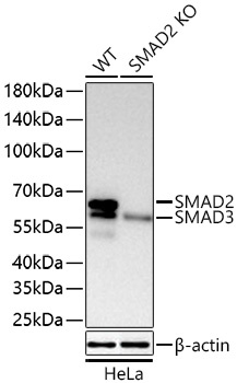
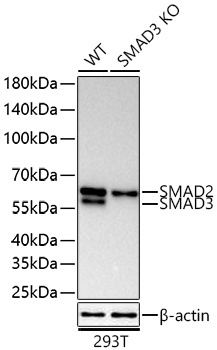
The [KO Validated] SMAD2/SMAD3 Rabbit mAb (CAB28421) is an unconjugated recombinant rabbit monoclonal antibody (clone ARC77767) developed for reliable detection of SMAD2 and SMAD3, the receptor-regulated SMAD proteins that transmit TGF-β and activin signals from the cell surface to the nucleus. Its specificity has been confirmed by knockout validation, with loss of the SMAD2 band in SMAD2 knockout HeLa cells and loss of the SMAD3 band in SMAD3 knockout 293T cells. SMAD2/SMAD3 are widely studied in cancer, fibrosis, immunology and developmental biology. This antibody is supplied as a kit with Bradford Reagent for optional total protein quantification and accurate sample normalization.
This antibody is validated for use in WB, IP, ChIP, ELISA applications and has demonstrated reactivity against Human, Mouse, Rat samples.
Synthetic peptide. This information is considered to be commercially sensitive.
Tested Applications:
WBIPChIPELISA
Recommended Dilution:
WB
1:6000 - 1:15000
IP
0.5 µg - 4 µg antibody for 200 µg - 400 µg extracts of whole cells
ChIP
2 µg antibody for 10 µg - 15 µg of chromatin
ELISA
Recommended starting concentration is 1 µg/mL. Please optimize the concentration based on your specific assay requirements. For high-ratio antibody dilutions (≥1:10000), a sequential dilution method is strongly recommended to ensure measurement accuracy.
SMAD2 and SMAD3 belong to the SMAD family of proteins, which are similar to the gene products of the Drosophila gene 'mothers against decapentaplegic' (Mad) and the C. elegans gene Sma. SMAD proteins are signal transducers and transcriptional modulators that mediate multiple signaling pathways. SMAD2 mediates the signal of transforming growth factor (TGF)-beta and thus regulates multiple cellular processes, such as cell proliferation, apoptosis and differentiation. It is recruited to the TGF-beta receptors through its interaction with the SMAD anchor for receptor activation (SARA) protein and, in response to TGF-beta signal, is phosphorylated by the TGF-beta receptors. Phosphorylation induces its dissociation from SARA and association with SMAD4, which is important for its translocation into the nucleus, where it binds to target promoters and forms a transcription repressor complex with other cofactors. SMAD2 can also be phosphorylated by activin type 1 receptor kinase and mediates the signal from activin. SMAD3 functions in the TGF-beta signaling pathway and transmits signals from the cell surface to the nucleus, regulating gene activity and cell proliferation. It forms a complex with other SMAD proteins and binds DNA, functioning both as a transcription factor and tumor suppressor. Mutations in SMAD3 are associated with aneurysms-osteoarthritis syndrome and Loeys-Dietz syndrome 3.
Purification Method
Affinity purification
Gene ID
4087/4088
UniProt
Q15796/P84022
Buffer Information
Store at -20°C. Avoid freeze / thaw cycles. Buffer: PBS with 0.09% sodium azide, 0.05% BSA, 50% glycerol, pH 7.3.
Bradford Reagent Storage
Store at room temperature for 1 year.
Protein Quantification (Optional)
To quantify total protein levels, use the Bradford Reagent included in this kit. Visit our Bradford Protein Assay Protocol to view the full protocol.
Western blot analysis of lysates from wild type (WT) and SMAD2 knockout (KO) HeLa cells using [KO Validated] SMAD2/SMAD3 Rabbit mAb (CAB28421) at 1:15000 dilution incubated overnight at 4°C. Secondary antibody: HRP-conjugated Goat anti-Rabbit IgG (H+L) at 1:10000 dilution. Lysates/proteins: 25 µg per lane. Blocking buffer: 3% nonfat dry milk in TBST. Detection: ECL Basic Kit. Exposure time: 45s.
Western blot analysis of lysates from wild type (WT) and SMAD3 knockout (KO) 293T cells using [KO Validated] SMAD2/SMAD3 Rabbit mAb (CAB28421) at 1:15000 dilution incubated overnight at 4°C. Secondary antibody: HRP-conjugated Goat anti-Rabbit IgG (H+L) at 1:10000 dilution. Lysates/proteins: 25 µg per lane. Blocking buffer: 3% nonfat dry milk in TBST. Detection: ECL Basic Kit. Exposure time: 45s.
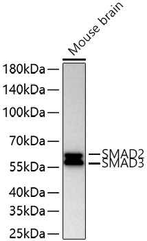
Western blot analysis of lysates from Mouse brain using [KO Validated] SMAD2/SMAD3 Rabbit mAb (CAB28421) at 1:15000 dilution incubated overnight at 4°C. Secondary antibody: HRP-conjugated Goat anti-Rabbit IgG (H+L) at 1:10000 dilution. Lysates/proteins: 25 µg per lane. Blocking buffer: 3% nonfat dry milk in TBST. Detection: ECL Basic Kit. Exposure time: 45s.
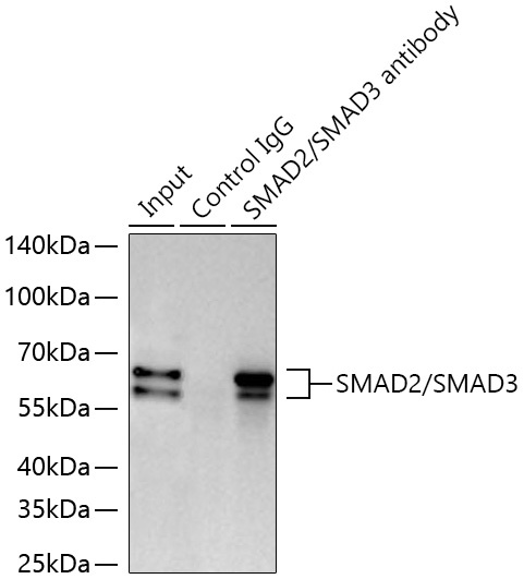
Immunoprecipitation of SMAD2/SMAD3 from 300 µg extracts of NIH/3T3 cells was performed using 0.5 µg of [KO Validated] SMAD2/SMAD3 Rabbit mAb (CAB28421). Rabbit Control IgG was used to precipitate the Control IgG sample. IP samples were eluted with 1x Laemmli Buffer. The Input lane represents 10% of the total input. Western blot analysis of immunoprecipitates was conducted using [KO Validated] SMAD2/SMAD3 Rabbit mAb (CAB28421) at a dilution of 1:6000.
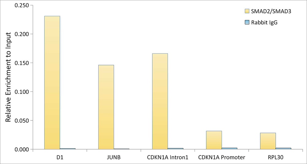
Chromatin immunoprecipitation was performed with 15 µg of cross-linked chromatin from A549 cells treated with TGF-β1 (20 ng/mL, 30 min), using 2 µg of [KO Validated] SMAD2/SMAD3 Rabbit mAb (CAB28421) and Rabbit IgG isotype control. The enrichment of immunoprecipitated DNA at different genomic loci was examined by quantitative PCR. The histogram compares the ratio of the immunoprecipitated DNA to the input at given loci.