Description
Myeloperoxidase (MPO) Peroxidation Activity Fluorometric Assay Kit
The Myeloperoxidase (MPO) Peroxidation Activity Fluorometric Assay Kit (MAES0004) measures MPO-specific peroxidase activity in serum, plasma, and animal tissue samples. A non-fluorescent probe is oxidized to a fluorescent product by peroxidase activity, and an MPO-specific inhibitor allows MPO activity to be separated from other peroxidases in the sample. Researchers use the kit to study neutrophil-derived oxidant activity and oxidative stress in research models. Research Use Only.
Myeloperoxidase (MPO) is a peroxidase, an enzyme class that uses hydrogen peroxide to oxidize substrates. In a biological sample, total peroxidase activity reflects several enzymes at once, so MPO-specific activity must be separated from that of other peroxidases. This kit addresses the problem by pairing a total peroxidase measurement with a parallel reaction containing an MPO-specific enzyme inhibitor. The difference between the two readings is reported as MPO peroxidation activity, expressed in units per liter (U/L) for serum and plasma and in units per gram of wet tissue weight for tissue homogenates.
Researchers measure MPO peroxidation activity in oxidative stress studies, the research area assigned to this kit. Publications that cite the kit include work in experimental intestinal inflammation in mice, drug-induced liver injury, lipopolysaccharide-induced acute lung injury, dextran sulfate sodium-induced colitis in mice, and aluminium chloride-induced vascular dementia in rats. These citations span inflammatory, hepatic, pulmonary, colonic, and neurovascular models. The kit is labeled for research use only and is not intended for clinical diagnosis.
The assay relies on a non-fluorescent probe that hydrogen peroxide oxidizes to a fluorescent product under peroxidase catalysis, so fluorescence intensity is proportional to total peroxidase activity. Control wells receive MPO inhibitor before the reaction working solution, which is prepared on the spot from buffer solution, probe, and substrate and protected from light. After the plate is mixed for 5 s, it is incubated at 37 °C for 10 min. Inhibitor is then added to sample and standard wells, and fluorescence is read at an excitation of 535 nm and an emission of 587 nm. The MPO-specific signal is the difference between the sample reading (F2) and the control reading (F1).
Serum and plasma are measured directly without extraction. If samples are not assayed on the day of collection, serum or plasma can be stored at -80 °C for up to one month. Animal tissue is washed in cold PBS (0.01 M, pH 7.4) and homogenized at 4 °C in buffer solution, with an initial recommendation of 20 mg tissue per 180 µL buffer. Homogenates are centrifuged at 10000 × g for 10 min to remove insoluble material, and the supernatant is kept on ice until assayed. Listed dilution factors cover porcine, rabbit, rat, mouse, and horse serum, mouse plasma, and 10% rat heart and lung homogenates.
Reported analytical sensitivity is 0.001 U/L, determined by adding two standard deviations to the mean of 20 zero-standard replicates and calculating the corresponding concentration. The detection range is 0.001 to 1.26 U/L. The product page lists intra-assay precision of 1.0% CV and inter-assay precision of 5.4% CV. The manual reports intra-assay CVs of 0.9% to 1.2% and inter-assay CVs of 4.8% to 6.2% across three human serum samples, assayed in replicate of 20 and 20 times, respectively. Standard curve values are provided for reference only, because fluorescence may vary with operator, pipetting, and temperature.
| Product name: | Myeloperoxidase (MPO) Peroxidation Activity Fluorometric Assay Kit |
| SKU: | MAES0004 |
| Sample types: | Serum, plasma, animal tissue |
| Detection method: | Fluorometric |
| Instrument: | Fluorescence microplate reader (Ex/Em = 535 nm/587 nm) |
| Sensitivity: | 0.001 U/L |
| Detection range: | 0.001-1.26 U/L |
| Assay time: | 20 min |
| Research area: | Oxidative stress; enzymes |
| Kit size: | 96 Assays |
| Detection method: | Fluorometric |
| Excitation/emission: | 535 nm/587 nm |
| Sensitivity (analytical): | 0.001 U/L |
| Detection range: | 0.001-1.26 U/L |
| Intra-assay CV: | 1.0% |
| Inter-assay CV: | 5.4% |
| Assay time: | 20 min |
| Sample volume per well: | 50 µL (sample or control) |
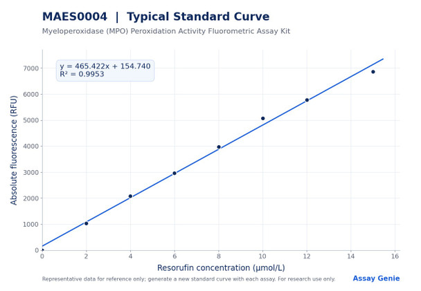
| Standard curve concentrations: | 0, 2, 4, 6, 8, 10, 12, 15 µmol/L resorufin |
| Reaction incubation: | 10 min at 37°C |
- Fluorometric detection of MPO peroxidase activity (Ex/Em = 535 nm/587 nm)
- MPO-specific inhibitor control distinguishes MPO from other peroxidases in the sample
- Detection range 0.001-1.26 U/L with analytical sensitivity of 0.001 U/L
- Intra-assay CV 1.0%; inter-assay CV 5.4%
- Approximately 20 min assay time plus a 10 min 37°C incubation
- Validated sample types: serum, plasma, and animal tissue homogenates
Under the catalysis of peroxidase, hydrogen peroxide oxidizes a non-fluorescent probe to a fluorescent product, and the fluorescence intensity is proportional to total peroxidase activity in the sample. The kit uses an MPO enzyme inhibitor to specifically inhibit MPO peroxidase activity in the sample. Measuring fluorescence with and without the inhibitor allows MPO activity to be distinguished from that of other peroxidases. MPO activity is calculated as F2 minus F1, where F1 is the inhibitor-containing control reading and F2 is the sample reading without inhibitor.
- Quantifying MPO-specific peroxidase activity in serum and plasma from research animals
- Measuring MPO peroxidase activity in animal tissue homogenates
- Studying neutrophil oxidant generation and host defense in experimental models
- Investigating MPO activity in models of cardiovascular disease, lung injury, and cancer
- Oxidative stress research using an MPO-selective readout
| Component | Quantity | Storage |
| Buffer Solution | 60 mL ×1 vial | -20°C, 12 months |
| Probe | 0.25 mL ×1 vial | -20°C, 12 months, protect from light |
| Substrate | 0.25 mL ×1 vial | -20°C, 12 months |
| Inhibitor (MPO inhibitor) | 1.2 mL ×1 vial | -20°C, 12 months |
| Resorufin Standard (25 µmol/L) | 1.5 mL ×1 vial | -20°C, 12 months, protect from light |
| Black microplate | 96 wells | No requirement |
| Plate sealer | 2 pieces | No requirement |
| Sample layout sheet | 1 piece | No requirement |
- Fluorescence microplate reader with excitation at 535 nm and emission at 587 nm
- Micropipettes and tips
- Vortex mixer
- Water bath or incubator at 37°C
- Centrifuge capable of 10000 ×g
- Homogenizer (Dounce-type) and ice bath for tissue samples
- Cold PBS (0.01 M, pH 7.4) for tissue washing; normal saline (0.9% NaCl) or PBS (0.01 M, pH 7.4) as diluent (see discrepancies)
- Protein assay reagents, user-supplied, to normalize tissue homogenate results
Serum and plasma
- Detect serum or plasma directly, without extraction.
- If samples are not detected on the same day, store serum or plasma at -80 °C for up to one month.
- Dilute samples using the recommended dilution factors: porcine serum 5-10; rabbit serum 3-5; rat serum 2-5; mouse serum 10-20; mouse plasma 30-50; horse serum 2-5.
- Use buffer solution as the diluent.
- If MPO peroxidation activity exceeds the detection range, dilute the sample to the optimal concentration for detection.
- For sample types not listed, run a pretest to confirm the dilution factor.
Animal tissue
- Harvest the amount of tissue needed for each assay (initial recommendation 20 mg).
- Wash tissue in cold PBS (0.01 M, pH 7.4).
- Homogenize 20 mg tissue in 180 µL buffer solution with a Dounce homogenizer at 4 °C.
- Centrifuge at 10000 × g for 10 min to remove insoluble material.
- Collect the supernatant and keep it on ice for detection.
- For 10% rat heart tissue homogenate and 10% rat lung tissue homogenate, use a dilution factor of 1.
- For other tissue types, run a pretest to confirm the dilution factor.
Reagent preparation
- Equilibrate all reagents to room temperature before use.
- Preheat the Buffer Solution at 37°C for 20 min; use only after it is completely clear.
- Prepare reaction working solution fresh for each well: 36 µL Buffer Solution, 2 µL Probe, and 2 µL Substrate (40 µL total). Prepare on the spot and protect from light.
- Prepare a fresh resorufin standard series by diluting the 25 µmol/L Resorufin Standard with Buffer Solution to 0, 2, 4, 6, 8, 10, 12, and 15 µmol/L. For each standard, add 0, 20, 40, 60, 80, 100, 120, or 150 µL of 25 µmol/L standard to 250, 230, 210, 190, 170, 150, 130, or 100 µL Buffer Solution, respectively. Discard working standards after use.
Sample preparation
- Serum and plasma: assay directly. If not assayed the same day, store at -80°C for up to one month.
- Tissue: collect the tissue amount needed for each assay (initial recommendation 20 mg). Wash in cold PBS (0.01 M, pH 7.4).
- Homogenize 20 mg tissue in 180 µL Buffer Solution with a Dounce homogenizer at 4°C.
- Centrifuge at 10000 ×g for 10 min. Collect the supernatant and keep it on ice until assayed.
- Determine protein concentration of the supernatant using a user-supplied protein assay.
Serum/plasma: MPO peroxidation activity (U/L) = (ΔF − b) ÷ a ÷ T × f. Tissue: MPO peroxidation activity (U/g tissue wet weight) = (ΔF − b) ÷ a ÷ T × f × V ÷ m × 1000
- ΔF is the absolute fluorescence value of the sample, F2 − F1, where F2 is the sample well reading and F1 is the control well reading.
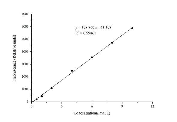
- a and b are the slope and intercept of the standard curve, y = ax + b, where y is the absolute fluorescence value and x is the resorufin concentration in µmol/L.
- T is the reaction time, 10 min.
- f is the dilution factor of the sample before testing. Use 1 for undiluted samples.
- m is the wet weight of the tissue sample, in g. This term applies to tissue samples only.
- V is the volume of buffer solution used for homogenization. This term applies to tissue samples only.
- 1000 is the conversion noted in the manual (1 U = 1000 mU).
- One unit is the amount of enzyme that produces 1 µmol of resorufin per minute at 37 °C, per liter of serum or plasma, or per gram of wet tissue weight for tissue.
Which sample types can be tested with this kit?
The kit detects MPO peroxidation activity in serum, plasma, and animal tissue samples. Tissue samples are homogenized and centrifuged, and the supernatant is used for detection.
How should reagents and samples be stored?
Reagents 1 through 5 are stored at -20 °C for 12 months, with the probe and resorufin standard also protected from light. Serum and plasma can be stored at -80 °C for up to one month if not tested on the day of collection.
What should I do if my results are above the detection range?
Dilute the sample to the optimal concentration for detection. The detection range is 0.001 to 1.26 U/L, and the recommended dilution factors are listed for each sample type.
What key points affect assay performance?
Prepare the reaction working solution and standard dilutions fresh, and keep them protected from light. Do not mix reagents from different kits, and centrifuge small reagent volumes before use.
What instrument settings are required?
Use a fluorescence microplate reader with excitation at 535 nm and emission at 587 nm, and a black microplate. Read the control well (F1) and the sample well (F2) after the 37 °C incubation.
How long does the assay take?
The reaction incubation is 37 °C for 10 min after mixing. The kit has an assay time of 20 min.