The Recombinant Phospho-eIF2α (Ser51) Monoclonal Antibody is a recombinant rabbit monoclonal antibody (clone C110105) against eIF2α (Ser51). The translation initiation factor EIF2 catalyzes the first regulated step of protein synthesis initiation, promoting the binding of the initiator tRNA to 40S ribosomal subunits. Binding occurs as a ternary complex of methionyl-tRNA, EIF2, and GTP. EIF2 is composed of 3 nonidentical subunits, the 36-kD EIF2-alpha subunit (EIF2S1), the 38-kD EIF2-beta subunit (EIF2S2; MIM 603908), and the 52-kD EIF2-gamma subunit (EIF2S3; MIM 300161). Produced by recombinant technology in Rabbit hosts and purified by Protein A affinity chromatography, this Rabbit IgG antibody is reactive against Human, Mouse and Rat samples.
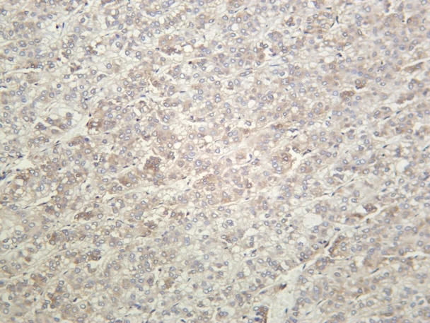
This antibody is suitable for WB, IHC, IF and ELISA applications. It has been validated by WB analysis of Hela, Hela was treated with Thapsigargin (300nM) for 30 minutes, IHC analysis of Human hepatocellular carcinoma and IF analysis of A549.
A synthetic phosphopeptide corresponding to residues around Ser51 of human Phospho-eIF2α.
Applications:
WBIHCIFELISA
Clonality:
Recombinant Monoclonal
Clone No:
C110105
Host:
Rabbit
Isotype:
Rabbit IgG
Target:
eIF2α (Ser51)
Conjugation:
Unconjugated
Observed MW:
36 kDa
Calculated MW:
36 kDa
Recommended Dilution:
IHC 1:100-1000; WB 1:2000-10000; IF 1:200-1000; ELISA 1:5000-20000
UniProt:
P05198, Q6ZWX6, P68101
Purification:
Protein A
Concentration:
1 mg/mL
Buffer:
0.2 µm filtered solution in PBS
Storage:
Store at -20°C valid for 12 months. Avoid freeze / thaw cycles.
Immunofluorescence analysis of A549. 1,primary (4℃ Antibody (red) was diluted at 1:200 overnight). 2, Goat Anti Rabbit IgG (H&L) - Alexa Fluor 594 Secondary antibody was diluted at 1:1000 (room temperature, 50min).3, DAPI (blue) 10min.
Human hepatocellular carcinoma was stained with anti- eIF2α (Phospho Ser51) Rabbit antibody
Various whole cell lysates were separated by 4-20% SDS- PAGE, and the membrane was blotted with anti-eIF2α (Phospho Ser51) antibody. The HRP-conjugated Goat anti-Rabbit IgG (H + L) antibody was used to detect the antibody. Lane 1: Hela Lane 2: Hela was treated with Thapsigargin (300nM) for 30 minutes Predicted band size: 36kDa Observed band size: 36kDa