Description
Xanthine Oxidase (XOD) Activity Assay Kit
The Assay Genie Xanthine Oxidase (XOD) Activity Assay Kit (MAES0265) measures XOD activity in serum, plasma, and animal tissue samples. XOD converts hypoxanthine to xanthine and generates superoxide, and the assay quantifies the resulting purplish red chromogenic product by absorbance at 550 nm. It is intended for Research Use Only and is used in oxidative stress research.
Xanthine oxidase (XOD) is an aerobic dehydrogenase found mainly in the milk, liver and spleen of mammals. It is an important enzyme in nucleic acid metabolism. During hypoxia, xanthine dehydrogenase rapidly forms xanthine oxidase, which plays an important role in free radical production. XOD catalyzes the conversion of hypoxanthine to xanthine while generating superoxide anion free radicals. Because of this reactive oxygen species output, the enzyme sits at the intersection of purine catabolism and oxidative stress.
Serum XOD activity is of interest in liver disease because the enzyme is released into serum earlier than SGPT when hepatocytes are injured, and its serum activity increases significantly. The source literature cites its value for identifying hepatocellular jaundice and for identifying obstructive jaundice. Because XOD contributes to free radical production during hypoxia, researchers also use this kit in oxidative stress research. The manual's example analysis applies the assay to mouse plasma and to rat and mouse liver and kidney tissue homogenates.
The assay is a colorimetric method. XOD converts hypoxanthine to xanthine and produces superoxide. In the presence of an electron acceptor and a chromogenic agent, a purplish red substance forms, and XOD activity is calculated from its production. The chromogenic agent 1 and agent 2 are mixed to form the chromogenic working solution, which is combined with buffer, substrate and enzymatic reagent. Each well receives 20 µL of standard or sample and 180 µL of working solution. After a 5 s mix, the first absorbance (A1) is read at 550 nm, the plate is incubated at 37°C for 25 min, and the second reading (A2) is taken. The OD change (ΔA550 = A2 - A1) is converted to activity using the standard curve.
Serum and plasma can be assayed directly. If they are not tested on the same day, they can be stored at -80°C for one month. For animal tissue, wash the tissue in cold PBS (0.01 M, pH 7.4) and homogenize 20 mg of tissue in 180 µL of normal saline (0.9% NaCl) with a Dounce homogenizer at 4°C. Centrifuge at 10000 × g for 10 min at 4°C to remove insoluble material, then keep the supernatant on ice for detection. Determine the protein concentration of the supernatant separately, because tissue results are normalized to protein. Ask researchers to run a pretest for sample types not listed in the manual.
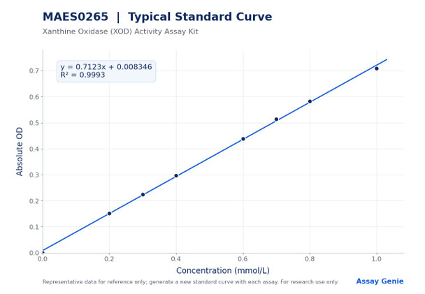
The analytical sensitivity is 0.067 U/L, and the detection range is 0.067 to 39.30 U/L. Intra-assay precision ranged from 2.6% to 3.4% CV across three human serum samples in replicates of 20, and inter-assay precision ranged from 9.0% to 10.4% CV over 20 runs by three operators. Recovery was 89%, 93% and 115% for three standards at low, middle and high concentrations, with an average recovery of 99% reported in the manual. The standard curve is provided for reference only, because optical density values vary with operator, pipetting and temperature.
| Product Name: | Xanthine Oxidase (XOD) Activity Assay Kit |
| SKU: | MAES0265 |
| Sample Type: | Serum, plasma, and animal tissue samples |
| Assay Type: | Enzyme activity |
| Synonyms: | XOD |
| Detection Method: | Colorimetric |
| Detection Instrument: | Microplate reader (540-560 nm; optimum 550 nm) |
| Assay Time: | 35 min |
| Sensitivity: | 0.067 U/L |
| Detection Range: | 0.067-39.30 U/L |
| Intra-assay CV: | 3% |
| Inter-assay CV: | 9.9% |
| Research Area: | Oxidative stress |
| Detection Method: | Colorimetric |
| Optimum Wavelength: | 550 nm (read range 540-560 nm) |
| Sensitivity: | 0.067 U/L |
| Detection Range: | 0.067-39.30 U/L |
| Intra-assay CV: | 3% |
| Inter-assay CV: | 9.9% |
| Sample Volume: | 20 µL |
| Assay Time: | 35 min |
- Colorimetric endpoint readout on a microplate reader (540-560 nm; optimum 550 nm)
- Detection range 0.067-39.30 U/L with a sensitivity of 0.067 U/L
- Assay time of 35 min with 20 µL sample input
- Validated for serum, plasma, and animal tissue homogenate samples
- Intra-assay CV of 3% and inter-assay CV of 9.9%
XOD catalyzes the conversion of hypoxanthine to xanthine while producing superoxide anion free radicals. In the presence of an electron acceptor and a chromogenic agent, a purplish red substance is generated. XOD activity is calculated from the rate of purplish red product formation, measured as the change in absorbance at 550 nm over 25 min at 37°C.
- Measuring XOD activity in mammalian serum and plasma samples
- Quantifying XOD activity in animal tissue homogenates (reported per gram protein)
- Investigating hypoxia-associated free radical production in research models
- Supporting oxidative stress research studies
| Component | Storage |
| Buffer Solution | -20°C, 12 months |
| Substrate Solution | -20°C, 12 months, protect from light |
| Enzymatic Reagent | -20°C, 12 months, protect from light |
| Chromogenic Agent 1 | -20°C, 12 months, protect from light |
| Chromogenic Agent 2 | -20°C, 12 months, protect from light |
| 1 mmol/L Standard Solution | -20°C, 12 months |
| Microplate | No requirement |
- Microplate reader (540-560 nm; optimum 550 nm)
- Incubator set to 37°C
- Normal saline (0.9% NaCl)
- Double-distilled water (for standard dilutions)
- PBS (0.01 M, pH 7.4) for tissue washing
- Homogenizer (Dounce type) and refrigerated centrifuge capable of 10000 × g
- Separate protein concentration assay for tissue samples
Serum/plasma
- Detect serum or plasma directly.
- If samples are not detected on the same day, store serum or plasma at -80°C for up to one month.
- Dilution factor: rat plasma and rat serum, 1 (no dilution required). For other sample types, run a pretest to confirm the dilution factor.
- Diluent: normal saline (0.9% NaCl) or PBS (0.01 M, pH 7.4).
Tissue
- Harvest the amount of tissue needed for each assay (initial recommendation: 20 mg).
- Wash the tissue in cold PBS (0.01 M, pH 7.4).
- Homogenize 20 mg tissue in 180 µL normal saline (0.9% NaCl) with a Dounce homogenizer at 4°C.
- Centrifuge at 10000 × g for 10 min at 4°C to remove insoluble material.
- Collect the supernatant and keep it on ice for detection.
- Determine the protein concentration of the supernatant separately for normalization.
- Dilution factor: 10% mouse kidney, rat liver, rat kidney and mouse liver tissue homogenates, 1 (no further dilution). For other sample types, run a pretest to confirm the dilution factor.
- Diluent: normal saline (0.9% NaCl) or PBS (0.01 M, pH 7.4).
Reagent preparation
- Equilibrate all reagents to room temperature before use.
- Prepare chromogenic working solution by diluting 20 µL of Chromogenic Agent 1 with 20 µL of Chromogenic Agent 2 and mixing well. Prepare fresh, protect from light, and use within 1 hour.
- Prepare working solution for each well: 147 µL Buffer Solution, 6.5 µL Substrate Solution, 6.5 µL Enzymatic Reagent, and 20 µL chromogenic working solution (180 µL total). Prepare fresh and protect from light.
- Prepare standards by diluting the 1 mmol/L Standard Solution with double-distilled water to 0, 0.2, 0.3, 0.4, 0.6, 0.7, 0.8, and 1.0 mmol/L. Prepare a fresh set for each assay and discard after use. For example, 0.4 mmol/L uses 80 µL standard and 120 µL water.
Sample preparation
- Serum and plasma: assay directly. If not assayed the same day, store at -80°C for up to one month.
- Tissue: wash tissue in cold PBS (0.01 M, pH 7.4). Homogenize 20 mg tissue in 180 µL normal saline (0.9% NaCl) with a Dounce homogenizer at 4°C.
- Centrifuge the homogenate at 10000 × g for 10 min at 4°C. Collect the supernatant and keep on ice.
- Determine the protein concentration of the supernatant with a separate protein assay for tissue calculations.
- Dilute samples in normal saline (0.9% NaCl). Recommended dilution factor is 1 for rat plasma, rat serum, and 10% mouse kidney, rat liver, rat kidney, and mouse liver tissue homogenates. Pretest other sample types to confirm dilution.
Assay procedure
- Add 20 µL of each standard to standard wells and 20 µL of sample to sample wells.
- Add 180 µL of working solution to each well.
- Mix for 5 s on the microplate reader, then read absorbance at 550 nm. Record as A1.
- Incubate at 37°C for 25 min, then read absorbance at 550 nm for sample and standard wells. Record as A2.
Serum/plasma: XOD activity (U/L) = (ΔA550 - b) ÷ a ÷ T × f × 1000 Tissue: XOD activity (U/g prot) = (ΔA550 - b) ÷ a ÷ T × f ÷ Cpr × 1000 ΔA550 = A2 - A1
- ΔA550: change in OD of the sample at 550 nm (A2 minus A1). A1 is read after mixing, and A2 is read after 25 min at 37°C.
- a: slope of the standard curve (y = ax + b), where y is the blank-subtracted absolute OD and x is the standard concentration in mmol/L.
- b: intercept of the standard curve (y = ax + b).
- T: reaction time in min, 25 min.
- f: dilution factor of the sample before testing. Multiply by f when the sample was diluted (for example, 1 for undiluted samples).
- Cpr: protein concentration of the tissue supernatant in g prot/L. Tissue results are expressed per gram of protein.
- 1000: converts 1 mmol/L to 1000 µmol/L, so the result is expressed as units per liter (serum/plasma) or units per gram of protein (tissue).
- Definition: 1 unit is the amount of XOD that hydrolyzes the substrate to produce 1 µmol H2O2 in 1 minute at 37°C.
How should serum and plasma samples be stored before testing?
Serum and plasma can be detected directly. If they are not tested on the same day, store them at -80°C for up to one month.
How should the kit reagents be stored, and how long are they stable?
All reagents should be stored at -20°C for 12 months, protected from light for the substrate, enzymatic reagent and chromogenic agents. Prepare the chromogenic working solution fresh and use it within 1 hour, and prepare the working solution on the spot and protect it from light.
What should I do if my sample reads outside the detection range?
The detection range is 0.067 to 39.30 U/L. If the concentration is not within this range, perform an extra dilution or concentration of the sample. Diluent is normal saline (0.9% NaCl) or PBS (0.01 M, pH 7.4).
What instrument settings and incubation conditions are required?
Read absorbance on a microplate reader at 540 to 560 nm, with 550 nm as the optimum wavelength. Mix for 5 s, read A1, incubate at 37°C for 25 min, and then read A2.
What is the total assay time?
The assay time is 35 min. Use 20 µL of sample or standard and 180 µL of working solution per well.
What should I check before testing a sample type that is not listed?
Run a pretest to confirm the dilution factor for any sample type not listed in the manual. The kit is for research use only and must not be used for clinical diagnosis.
| Storage: | -20°C |
| Shelf Life: | 12 months |
| Light Protection: | Store reagents with shading light |
Notes
- For Research Use Only. Not for clinical diagnosis or other diagnostic use.
- Do not mix reagents from different kits.
- Centrifuge small-volume reagents before use to ensure sufficient volume.
- Protect chromogenic working solution and working solution from light, and use the chromogenic working solution within 1 hour.
- Prepare fresh standards for each assay and discard working standard dilutions after use.
- If a sample is outside the detection range, dilute or concentrate it and re-assay.
- Run a pretest for sample types not listed in the protocol.
- Wear a lab coat and latex gloves during handling.