Description
Xanthine Oxidase (XOD) Activity Fluorometric Assay Kit
The Xanthine Oxidase (XOD) Activity Fluorometric Assay Kit (MAES0006) measures XOD enzyme activity in serum, plasma, and animal tissue homogenates. XOD generates hydrogen peroxide from hypoxanthine, and the peroxide is detected with a fluorogenic probe in a 96-well format. Researchers use the kit to quantify XOD activity in oxidative stress and enzyme studies.
Xanthine oxidase (XOD) is an enzyme that oxidizes hypoxanthine to produce xanthine and a superoxide anion. In the system, the superoxide anion quickly converts to hydrogen peroxide, so XOD activity can be tracked through the peroxide it generates. This kit measures XOD activity in serum, plasma, and animal tissue samples, and the product is listed under the oxidative stress research area.
Researchers use this kit to quantify XOD activity in serum, plasma, and tissue homogenates, which is the core readout for studies in the oxidative stress research area. Because the assay detects hydrogen peroxide generated by the enzyme, it links XOD turnover to a peroxide-producing pathway. The kit is labeled for research use only and is not intended for clinical diagnosis. The sources do not describe specific disease models, so applications are limited to the sample types and research area named here.
The assay relies on a peroxidase-coupled fluorometric reaction. Hydrogen peroxide produced from hypoxanthine oxidation oxidizes a non-fluorescent probe to a fluorescent product, and the fluorescence signal is read on a fluorescence microplate reader at Ex/Em = 535 nm/587 nm. Each sample is run with a sample working solution containing substrate, probe, and enzyme application solution, plus a control working solution that lacks substrate. The first reading is taken, the plate is held at room temperature in the dark for 10 min, and a second reading is taken. The difference between the two readings, corrected for the control well, is used for calculation.
Serum and plasma are tested directly without pretreatment. Animal tissue is washed in cold PBS (0.01 M, pH 7.4), homogenized at 4°C in buffer solution, and centrifuged to remove insoluble material, and the supernatant is kept on ice. Tissue results are expressed per gram of protein, so the protein concentration of the supernatant must also be determined. Sample dilution is required for several matrices, with listed factors of 1 for mouse serum, rat plasma, horse serum, and human plasma, 3-5 for dog serum, and 5-10 for 10% rat kidney, mouse heart, and rat lung homogenates. For other sample types, a pretest is recommended to confirm the dilution factor.
Precision was evaluated with three human serum samples. Intra-assay precision, based on 20 replicates per sample, gave a coefficient of variation of 3.3% to 4.8% at mean levels of 0.10 to 1.00 U/L. Inter-assay precision, based on 20 runs in duplicate by three operators, gave a coefficient of variation of 8.5% to 10.0% across the same sample levels. The analytical sensitivity is 0.01 U/L, and the detection range is 0.01 to 1.2 U/L. Recovery data are not provided in the source material.
| Product Name: | Xanthine Oxidase (XOD) Activity Fluorometric Assay Kit |
| SKU: | MAES0006 |
| Size: | 96 Assays |
| Sample Type: | Serum, plasma, animal tissue homogenate |
| Detection Method: | Fluorometric |
| Instrument: | Fluorescence microplate reader (Ex/Em 535 nm/587 nm) |
| Assay Time: | 25 min |
| Research Area: | Enzymes, oxidative stress |
| Research Use Only: | Yes. Not for diagnostic or therapeutic use |
| Sensitivity: | 0.01 U/L |
| Detection Range: | 0.01–1.2 U/L |
| Intra-assay CV: | 4.1% |
| Inter-assay CV: | 9.0% |
| Excitation/Emission: | 535 nm/587 nm |
| Sample Volume: | 50 µL per well |
| Assay Time: | 25 min |
| Sample Types: | Serum, plasma, animal tissue homogenate |
- Fluorometric detection read at Ex/Em 535 nm/587 nm on a fluorescence microplate reader
- Detection range 0.01–1.2 U/L with analytical sensitivity of 0.01 U/L
- Validated for serum, plasma, and animal tissue homogenate samples
- Paired sample and control wells correct for background fluorescence
- Assay time of 25 min after reagent preparation
- 96-test kit supplied with black microplate and plate sealers
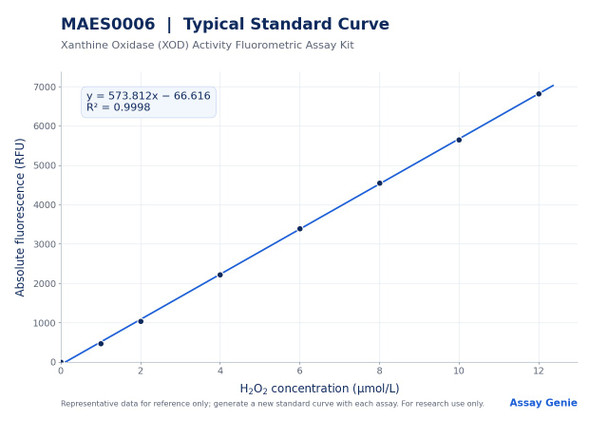
Hypoxanthine is oxidized by xanthine oxidase (XOD) to produce xanthine and superoxide anion. The superoxide rapidly converts to hydrogen peroxide (H2O2) in the system. In the presence of peroxidase, H2O2 oxidizes a non-fluorescent probe to a fluorescent product. Fluorescence is measured at Ex/Em 535 nm/587 nm, and XOD activity is calculated from the fluorescence change against an H2O2 standard curve.
- Measurement of XOD activity in mouse, rat, dog, and horse serum and plasma
- XOD activity profiling in rat and mouse heart, lung, and kidney tissue homogenates
- Oxidative stress studies involving hydrogen peroxide generation by XOD
- Purine metabolism and uric acid pathway research
- Liver-related enzyme studies in animal models
| Component | Quantity | Storage |
| Reagent 1: Buffer Solution | 60 mL×1 vial | -20°C, 12 months |
| Reagent 2: Probe Solution | 0.3 mL×1 vial | -20°C, 12 months, protect from light |
| Reagent 3: Enzyme Powder | 1 vial | -20°C, 12 months |
| Reagent 4: Substrate | 4 mL×1 vial | -20°C, 12 months, protect from light |
| Reagent 5: 2 mmol/L H2O2 Standard Solution | 1.5 mL×1 vial | -20°C, 12 months |
| Black Microplate | 96 wells×1 | No requirement |
| Plate Sealer | 2 pieces | No requirement |
| Sample Layout Sheet | 1 piece | No requirement |
- Fluorescence microplate reader with Ex/Em 535 nm/587 nm capability
- Vortex mixer
- Centrifuge (including microcentrifuge capable of 10000×g)
- Double-distilled water
- Normal saline (0.9% NaCl) or PBS (0.01 M, pH 7.4) for sample dilution
- Cold PBS (0.01 M, pH 7.4) for tissue washing
- Dounce homogenizer
- Ice bath and 4°C refrigeration for tissue homogenization
- Protein quantification method for tissue supernatant (not included in kit)
- Pipettes and tips, graph or spreadsheet software for standard curve fitting
Serum and plasma
- Detect serum and plasma samples directly without pretreatment.
- If samples are not tested on the same day, store serum or plasma at -80°C for up to one month.
- Dilute samples according to the recommended factors: mouse serum 1; dog serum 3-5; rat plasma 1; horse serum 1; human plasma 1.
- For sample types not listed, perform a pretest to confirm the dilution factor.
- Dilute with normal saline (0.9% NaCl) or PBS (0.01 M, pH 7.4) as listed in the product information.
Animal tissue (homogenate)
- Harvest the amount of tissue needed for each assay; the initial recommendation is 20 mg.
- Wash the tissue in cold PBS (0.01 M, pH 7.4).
- Homogenize 20 mg of tissue in 180 µL of buffer solution with a Dounce homogenizer at 4°C.
- Centrifuge at 10000×g for 10 min to remove insoluble material.
- Collect the supernatant and keep it on ice for detection.
- Determine the protein concentration of the supernatant, which is required for expressing activity per gram of protein.
- Dilute the 10% rat kidney, 10% mouse heart, and 10% rat lung homogenates 5-10 fold before testing.
Reagent preparation
- Equilibrate all reagents to room temperature before use. Centrifuge small-volume reagents before opening.
- Enzyme application solution: dissolve one vial of enzyme powder in 300 µL of buffer solution and mix well. Store aliquots at -20°C for up to 30 days and avoid repeated freeze/thaw cycles.
- Sample working solution (50 µL per well): mix 46 µL substrate, 2 µL probe solution, and 2 µL enzyme application solution. Prepare on the spot and protect from light.
- Control working solution (50 µL per well): mix 46 µL buffer solution, 2 µL probe solution, and 2 µL enzyme application solution. Prepare on the spot and protect from light.
- 20 µmol/L H2O2 standard: add 5 µL of 2 mmol/L H2O2 standard solution to 495 µL double-distilled water and mix. Prepare on the spot.
Standard curve
- Prepare a fresh set of standards each time and discard working dilutions after use.
- Dilute the 20 µmol/L H2O2 standard with double-distilled water to 0, 1, 2, 4, 6, 8, 10, and 12 µmol/L (for example, 0 to 120 µL of 20 µmol/L standard plus water to a final 200 µL).
Sample preparation
- Serum and plasma: assay directly. If not assayed the same day, store at -80°C for up to one month.
- Tissue: wash tissue in cold PBS (0.01 M, pH 7.4). Homogenize 20 mg tissue in 180 µL buffer solution with a Dounce homogenizer at 4°C. Centrifuge at 10000×g for 10 min and keep the supernatant on ice.
- Determine protein concentration of the tissue supernatant for tissue activity normalization.
- Dilute samples as needed (for example, mouse serum 1; dog serum 3–5; rat plasma 1; horse serum 1; human plasma 1; 10% rat kidney, mouse heart, or rat lung homogenate 5–10). Diluent is buffer solution or normal saline/PBS. Pretest other sample types.
Assay
- Add 50 µL of standards to standard wells, 50 µL of sample to sample wells, and 50 µL of sample to control wells.
- Add 50 µL of sample working solution to standard and sample wells. Add 50 µL of control working solution to control wells.
- Mix for 5 s on the plate reader and stand at room temperature for 2 min.
- Read fluorescence at Ex/Em 535 nm/587 nm (F1). Stand at room temperature protected from light for 10 min, then read again under the same conditions (F2).
- Calculate ΔF(sample) = F2(sample) − F1(sample) and ΔF(control) = F2(control) − F1(control). Absolute value ΔF = ΔF(sample) − ΔF(control). Standard wells show no change over time; use F2 of standards for the curve.
Serum/plasma: XOD activity (U/L) = (ΔF - b) ÷ a ÷ T × f; Tissue: XOD activity (U/gprot) = (ΔF - b) ÷ a ÷ T × f ÷ C_pr
- ΔF is the absolute fluorescence value of the sample, calculated as F_sample − F_control, where F_sample = F2(sample) − F1(sample) and F_control = F2(control) − F1(control).
- a and b are the slope and intercept of the standard curve y = ax + b, where y is the absolute fluorescence value (ΔF) of the standards and x is the H2O2 concentration in µmol/L.
- T is the reaction time in minutes, which is 10 min.
- f is the dilution factor of the sample before testing; use 1 for undiluted samples.
- C_pr is the protein concentration of the sample in gprot/L, used only for tissue samples.
- One unit of XOD is the amount that catalyzes the production of 1 µmol H2O2 per minute at 25°C; serum and plasma results are expressed in U/L, and tissue results are expressed in U/gprot.
Which sample types can be tested with this kit?
The kit measures XOD activity in serum, plasma, and animal tissue samples. Tissue samples are prepared as homogenates and require protein concentration measurement for calculation.
How should samples and reagents be stored?
The kit components are stored at -20°C for 12 months, with the probe and substrate protected from light. Aliquot the enzyme application solution, which is stable for 30 days at -20°C, and avoid repeated freeze/thaw cycles. Serum and plasma not tested on the same day can be stored at -80°C for one month.
What should I do if my results fall outside the detection range?
The detection range is 0.01 to 1.2 U/L. If the concentration is not within this range, an extra dilution or concentration of the sample should be performed.
What are the key points for a reliable assay?
Keep the reaction time accurate and limit each batch to fewer than 20 samples. Prepare the sample and control working solutions fresh and protect them from light, and prepare standards fresh for each run. Run a pretest for any sample type not listed in the instructions.
What instrument settings are required?
Use a fluorescence microplate reader set to an excitation wavelength of 535 nm and an emission wavelength of 587 nm. Use a black microplate.
How long does the assay take?
The assay time is 25 min. After mixing and a 2 min room temperature hold, the first reading is taken, followed by a 10 min incubation in the dark and a second reading.
| Storage: | -20°C, protect probe and substrate from light |
| Valid Period (Shelf Life): | 12 months |
| Shipping: | Gel pack |
| Prepared Enzyme Solution: | Store aliquots at -20°C for up to 30 days; avoid repeated freeze/thaw |
| Sample Working Solutions: | Prepare on the spot; protect from light |
| Serum/Plasma Samples: | Assay same day; store at -80°C for up to one month |
Notes
- For Research Use Only. Not for clinical diagnosis or other diagnostic use.
- Protect probe solution, substrate, and working solutions from light.
- Store reagents strictly at -20°C. Do not mix reagents from different kits.
- Avoid repeated freeze/thaw cycles of the enzyme application solution.
- Keep the reaction time accurate; the 10 min reaction interval is used in the calculation.
- Limit each batch to fewer than 20 samples.
- Prepare standards fresh and discard working dilutions after use.
- If a sample falls outside the detection range, dilute or concentrate it and re-assay.
- Run a pretest for sample types not listed in the protocol.