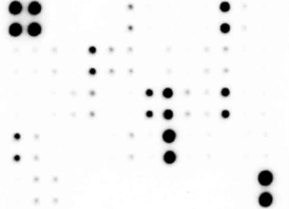
The Human/Mouse Akt Pathway Phosphorylation Array (SARB0017) is a cutting-edge tool for studying signaling pathways involved in cell growth, survival, and proliferation. This array allows for the simultaneous detection of phosphorylation levels of 18 key proteins in the Akt pathway, providing researchers with valuable insights into cellular signaling mechanisms.With the ability to analyze both human and mouse samples, this array is versatile and suitable for a wide range of experiments. Whether studying cancer biology, cell metabolism, or drug responses, the Human/Mouse Akt Pathway Phosphorylation Array offers a comprehensive view of Akt pathway activation in various biological contexts.
Designed for easy and efficient analysis, this array enables high-throughput screening of multiple phosphorylation events in a single experiment. By identifying changes in protein phosphorylation patterns, researchers can unravel the complex network of signaling cascades that regulate cellular functions. Gain a deeper understanding of Akt pathway activation with the Human/Mouse Akt Pathway Phosphorylation Array and uncover new possibilities for therapeutic interventions in diseases such as cancer and metabolic disorders.
Multiplexed Protein Detection; Detection of Relative Protein Expression; Detecting Patterns of Cytokine Expression; Biomarker/ Key Factor Screening; Identifying Key Factors; Confirming a Biological Process
Storage/Stability:
For best results, store the entire kit frozen at -20°C upon arrival. Stored frozen, the kit will be stable for at least 6 months which is the duration of the product warranty period. Once thawed, store array membranes and 1X Blocking Buffer at -20°C and all other reagents undiluted at 4°C for no more than 3 months.
Hover over target for more information
Akt (P-Ser473) (PKB (P-Ser473))
Gene Symbol: AKT1 PKB RAC
Gene ID: 207
Accession #: P31749
AMPKa (P-Thr172)
Gene Symbol: PRKAA1 AMPK1
Gene ID: 5562
Accession #: Q13131
BAD (P-Ser112)
Gene Symbol: BAD BBC6 BCL2L8
Gene ID: 572
Accession #: Q92934
4E-BP1 (P-Thr36)
Gene Symbol: EIF4EBP1
Gene ID: 1978
Accession #: Q13541
ERK1 (P-T202/Y204)/ERK2 (P-Y185/Y187)
Gene Symbol: MAPK1 ERK2 PRKM1 PRKM2
Gene ID: 5594
Accession #: P28482
GSK3a (P-Ser21)
Gene Symbol: GSK3A
Gene ID: 2931
Accession #: P49840
GSK3b (P-Ser9)
Gene Symbol: GSK3B
Gene ID: 2932
Accession #: P49841
mTOR (P-Ser2448)
Gene Symbol: MTOR FRAP FRAP1 FRAP2 RAFT1 RAPT1
Gene ID: 2475
Accession #: P42345
p27 (P-Thr198)
Gene Symbol: PSMD9
Gene ID: 5715
Accession #: O00233
P53 (P-Ser15)
Gene Symbol: TP53 P53
Gene ID: 6198
Accession #: P23443
P70S6K (P-Thr421/Ser424)
Gene Symbol: RPS6KB1 STK14A
Gene ID: 6198
Accession #: P23443
PDK1 (P-Ser241)
Gene Symbol: PDK1 PDHK1
Gene ID: 5170
Accession #: O15530
PRAS40 (P-Thr246)
Gene Symbol: AKT1S1 PRAS40
Gene ID: 84335
Accession #: Q96B36
PTEN (P-Ser380)
Gene Symbol: N/A
Gene ID: N/A
Accession #: P60484; O08586; O54857
Raf-1 (Ser301)
Gene Symbol: RAF1 RAF
Gene ID: 5894
Accession #: P04049
RPS6 (P-Ser235/Ser236)
Gene Symbol: RPS6 OK/SW-cl.2
Gene ID: 6194
Accession #: P62753
RSK1 (P-Ser380)
Gene Symbol: RPS6KA1 MAPKAPK1A RSK1
Gene ID: 6195
Accession #: Q15418
RSK2 (P-Ser386)
Gene Symbol: RPS6KA3 ISPK1 MAPKAPK1B RSK2
Gene ID: 10432
Accession #: P51812
Easy to use
No specialized equipment needed
Compatible with nearly any liquid sample
Proven technology
Highly sensitive
Sandwich ELISA specificity
Higher density than ELISA, Western blot or bead-based multiplex
Human and Mouse AKT Pathway Phosphorylation Antibody Array C1 Membranes
Blocking Buffer
Detection Antibody Cocktail
1,000X HRP-Anti-Rabbit IgG Concentrate
20X Wash Buffer I Concentrate
20X Wash Buffer II Concentate
2X Cell Lysis Buffer Concentrate
Detection Buffer C
Detection Buffer D
8-Well Incubation Tray
Protease Inhibitor Cocktail
100x Phosphatase Inhibitor Cocktail I
Phosphatase Inhibitor Cocktail II
Plastic Sheets
Array Map Template
Other Materials Required
Pipettors, pipet tips and other common lab consumables
Orbital shaker or oscillating rocker
Tissue Paper, blotting paper or chromatography paper
Adhesive tape or Saran Wrap
Distilled or de-ionized water
A chemiluminescent blot documentation system , X-ray Film and a suitable film processor, or another chemiluminescent detection system.
1.
Block membranes
2.
Incubate with Sample
3.
Incubate with Detection Antibody Cocktail
4.
Incubate with HRP-Conjugated anti-IgG
5.
Incubate with Detection Buffers
6.
Image with chemiluminescent imaging system
7.
Perform densitometry and analysis
De et al.
Hotspot mutations in HER2 interfaces destabilize structure, causing breast cancer treatment failure