


Description
TUNEL 1-Step In Situ Apoptosis Kit (FITC)
The TUNEL 1-Step In Situ Apoptosis Kit (FITC) (AKES071) is a fluorometric assay for detecting DNA fragmentation in apoptotic cells in situ. Terminal Deoxynucleotidyl Transferase (TdT) adds fluorescein-labeled dUTP to exposed 3'-OH ends of DNA breaks. Labeled cells are read directly by fluorescence microscopy with a FITC filter set. The kit is for research use only.
Apoptosis is a programmed cell death process in which specific DNA endonucleases are activated and cut genomic DNA between nucleosomes. The DNA of apoptotic cells is cleaved into multimers of 180 to 200 bp fragments, corresponding to oligonucleosomal units, and typically migrates as a ladder of 180 to 200 bp on an agarose gel. The exposed 3'-OH ends generated by this cleavage are the molecular feature this kit targets, so apoptotic cells can be identified in situ rather than by gel analysis of extracted DNA.
This kit is intended for detecting apoptotic cells directly in tissue and cell preparations, so the spatial location of labeled cells is retained within the specimen. It supports paraffin-embedded and frozen tissue sections as well as cell slides and cell smears, which allows researchers to compare apoptotic signal with tissue morphology. The kit is for research use only and is not intended for diagnostic use.
The 3'-OH ends are labeled by terminal deoxynucleotidyl transferase (TdT), which incorporates fluorescein-labeled dUTP into the exposed DNA ends. The labeling working solution is prepared fresh by combining TdT Equilibration Buffer, FITC Labeling Solution and TdT Enzyme, then applied at 50 µL per sample and incubated for 60 min at 37 °C in a humidified chamber with shading light. Labeled sites are read with a fluorescence microscope using a FITC filter set (excitation/emission 490/520 nm). Nuclei may be counterstained with DAPI before mounting in an anti-fluorescence quenching agent.
Suitable specimens are cell slides, cell smears, paraffin sections and frozen sections. Fixation in 4% paraformaldehyde in PBS is a critical step, because unfixed cells may lose smaller DNA fragments and produce lower signals. Permeabilization uses 0.2% Triton X-100 in PBS. Samples should be kept moist throughout the procedure, and labeled slides should be observed as soon as possible or stored at 4 °C protected from light.
| Application: | DNA fragmentation detection (apoptosis) |
| Detection method: | Fluorometric |
| Sample types: | Paraffin section, frozen section, cell slide |
| Assay time: | 3 hours |
| Detection instrument: | Fluorescence microscope |
| Dye type: | FITC |
| Ex/Em (nm): | 490/520 |
| Channel set: | FITC |
| Filter set: | FITC |
- One-step TdT-mediated labeling of exposed 3'-OH DNA ends with fluorescein-labeled dUTP
- FITC label, Ex/Em 490/520 nm, read on a FITC filter set
- Applicable to paraffin sections, frozen sections, and cell slides
- Assay time of 3 hours
- Results observed directly by fluorescence microscopy, with no separate detection instrument required
- Shelf life of 12 months at -20°C
Apoptotic DNA endonucleases generate 3'-OH ends in genomic DNA. Terminal Deoxynucleotidyl Transferase (TdT) catalyzes the incorporation of fluorescein-labeled dUTP onto these exposed 3'-OH ends. Incorporated fluorescein is then visualized by fluorescence microscopy with a FITC filter set (Ex/Em 490/520 nm).
- In situ detection of apoptosis in paraffin-embedded and frozen tissue sections
- Detection of apoptotic DNA fragmentation in cell slides and cell smears
- Fluorescence microscopy analysis of chemically induced apoptosis in cultured cells
- Assessment of DNA strand breaks in tissue, with DNase I-treated sections used as a positive control
| Component | Storage |
| TdT Equilibration Buffer | -20°C |
| TdT Enzyme | -20°C |
| Proteinase K (100×) | -20°C |
| Labeling Solution (FITC) | -20°C, protect from light |
| DNase I (20 U/µL) | -20°C |
| DNase I Buffer (10×) | -20°C |
| DAPI Reagent (25 µg/mL) | 2-8°C or -20°C, protect from light |
- 4% paraformaldehyde (fixative)
- 0.2% Triton X-100 in PBS (permeabilization)
- PBS
- ddH2O
- Xylene and ethanol series (paraffin sections)
- Mounting medium with anti-fluorescence quencher
- Fluorescence microscope with FITC filter set
- Humidified chamber and 37°C incubator
- Absorbent filter paper
- DAPI for nuclear counterstain (optional)
Cell slides
- Wash the slides with PBS for 1 time.
- Immerse the cell slides in fixative buffer (4% paraformaldehyde in PBS) at RT for 15 to 20 min or at 4 °C for 1 to 2 h.
- Wash the slides with PBS for 3 times, 5 min each time.
- Permeabilize with 0.2% Triton X-100 in PBS.
- Wash the slides with PBS for 3 times, 5 min each time.
- Proceed to the equilibration and labeling steps.
Cell smears
- Collect the cells and resuspend them in PBS.
- Add an equal volume of fixative buffer (4% paraformaldehyde in PBS) and fix at RT for 15 to 20 min or at 4 °C for 1 to 2 h.
- Centrifuge at 600 × g for 5 min.
- Resuspend the cells in PBS and spread 25 to 50 µL of cell suspension on slides; allow to dry.
- Wash the slides with PBS for 3 times, 5 min each time.
- Permeabilize with 0.2% Triton X-100 in PBS, then wash with PBS for 3 times, 5 min each time.
- Proceed to the equilibration and labeling steps.
Paraffin sections
- Deparaffinize and hydrate the sections by conventional methods: immerse slides in xylene for 2 times, 10 min each, then hydrate through a graded ethanol series (100%, 95%, 90%, 80%, 75%), 3 min each step.
- Wash the slides with PBS for 3 times, 5 min each time.
- Absorb moisture around the tissue with filter paper. Add 100 µL of 1× Proteinase K working solution (1 µL Proteinase K (100×) in 99 µL PBS, prepared fresh) to each sample and incubate at 37 °C for 20 min.
- Wash the slides with PBS for 3 times, 5 min each time.
- Proceed to the equilibration and labeling steps. Incubation time may vary with tissue type and species; a preliminary experiment is recommended to confirm it.
Frozen sections
- Remove the frozen sections and equilibrate to room temperature.
- Immerse the slides in fixative buffer (4% paraformaldehyde in PBS) and incubate at RT (15 to 25 °C) for 30 min.
- Wash the slides with PBS for 2 times, 5 min each time.
- Place the slides in permeabilization buffer (0.2% Triton X-100 in PBS) and incubate at 37 °C for 10 min.
- Wash the slides with PBS for 3 times, 5 min each time.
- Add 100 µL of 1× Proteinase K working solution to each sample and incubate at 37 °C for 10 to 20 min. Incubation time may vary with tissue type and species; a preliminary experiment is recommended to confirm it.
- Wash the slides with PBS for 3 times, 5 min each time.
- Proceed to the equilibration and labeling steps.
Cell slides and cell smears
- Wash slides with PBS once. For cell smears, resuspend cells in PBS, add an equal volume of 4% paraformaldehyde, and fix at room temperature for 15~20 min or at 4°C for 1~2 h; centrifuge at 600×g for 5 min before spreading on slides.
- Fix cells in 4% paraformaldehyde at room temperature for 15~20 min or at 4°C for 1~2 h.
- Wash slides with PBS 3 times, 5 min each.
- Permeabilize with 0.2% Triton X-100 in PBS, then wash with PBS 3 times, 5 min each.
Paraffin sections
- Deparaffinize and hydrate sections through xylene and a graded ethanol series (100%, 95%, 90%, 80%, 75%, 3 min each step).
- Fix sections in 4% paraformaldehyde, then wash with PBS.
- Permeabilize with 0.2% Triton X-100, then wash with PBS.
- Absorb moisture around the section with filter paper and apply Proteinase K working solution; incubate at 37°C, with the time optimized for the tissue and species.
- Wash with PBS 3 times, 5 min each.
Frozen sections
- Equilibrate frozen sections to room temperature and fix in 4% paraformaldehyde at room temperature (15~25°C) for 30 min.
- Wash with PBS 2 times, 5 min each.
- Permeabilize with 0.2% Triton X-100 at 37°C for 10 min.
- Apply Proteinase K working solution at 37°C for 10~20 min, with the time optimized for the tissue and species.
- Wash with PBS 3 times, 5 min each.
Labeling and detection
- Blot around the sample, add 100 µL TdT equilibration buffer, and incubate at 37°C for 10~30 min.
- Blot around the sample without allowing it to dry. Add 50 µL labeling working solution (TdT enzyme plus labeling solution) and incubate at 37°C for 60 min in a humidified chamber, protected from light.
- Wash with PBS 3 times, 5 min each.
- Optional: add DAPI working solution and incubate at room temperature for 5 min in the dark; wash with PBS 4 times, 5 min each.
- Seal slides with anti-fluorescence quenching mounting medium.
- Analyze with a fluorescence microscope using a FITC filter set (Ex/Em 490/520 nm). Observe results promptly.
Which sample types are supported?
The kit supports cell slides, cell smears, paraffin sections and frozen sections. Results are viewed directly with a fluorescence microscope.
How should the kit be stored, and how long is it stable?
Store the kit at -20 °C for 12 months, protected from light. The kit ships on an ice bag.
What should I do if the signal is weak or the background is high?
For weak signal, extend the labeling incubation; the manual notes that up to 4 hours at 37 °C may be required for some systems. For high background, dilute TdT Equilibration Buffer 1:2 to 1:10 and fix samples immediately after collection.
Which filter set and wavelengths should I use?
Use a FITC filter set with excitation/emission of 490/520 nm. If DAPI counterstaining is used, the DAPI filter set (350/470 nm) is appropriate.
What controls should be included, and how long does the assay take?
Set up a positive control, treated with DNase I (200 U/mL) to cut DNA and generate exposed 3'-OH ends, and a negative control in which TdT Enzyme is omitted from the labeling solution. The assay time is 3 hours.
| Storage: | -20°C, protected from light; store for 12 months |
| Shipping: | Ice pack |
| Shelf life: | 12 months |
Cautions
- For research use only. Not for diagnostic or therapeutic use.
- Store TdT enzyme strictly at -20°C; return it to -20°C immediately after use.
- Avoid repeated freeze-thaw cycles of labeling solution and TdT enzyme.
- Do not vortex TdT enzyme, labeling solution, or DNase I; mix by gentle pipetting.
- Keep samples moist throughout the procedure to prevent failure from dry slides.
- Use neutral, non-acidic fixatives; acidic fixatives and fixation with ethanol or methanol can impair labeling.
- Protect slides from light during labeling and storage, and observe results as soon as possible.
- Incubation times for Proteinase K and labeling may need optimization for different tissues or species.
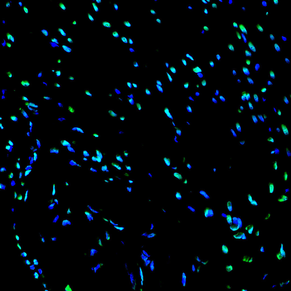 | Paraffin embedded rat heart was treated with DNase I to fragment the DNA. DNA strand breaks showed intense fluorescent staining in DNase I treated sample (green). The cells were counterstained with DAPI (blue). This photo was taken by confocal microscope. |
 | Paraffin embedded mouse liver was treated with DNase I to fragment the DNA. DNA strand breaks showed intense fluorescent staining in DNase I treated sample (green). The cells were counterstained with DAPI (blue).This photo was taken by confocal microscope |
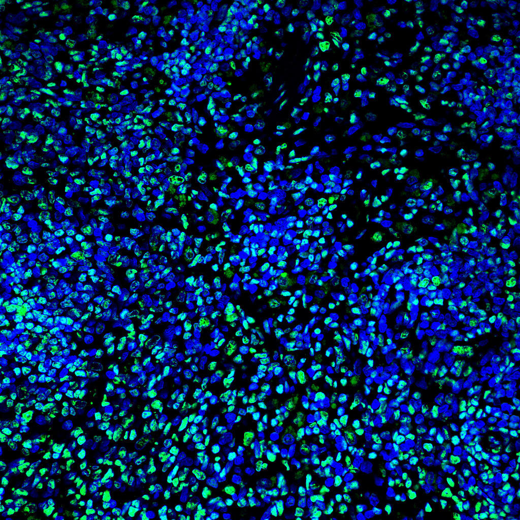 | Paraffin embedded mouse spleen was treated with DNase I to fragment the DNA. DNA strand breaks showed intense fluorescent staining in DNase I treated sample (green). The cells were counterstained with DAPI (blue). This photo was taken by confocal microscope. |
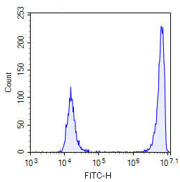 | Mixed samples of normal mouse spleen cells and DNase I treated mouse spleen cells were stained. |
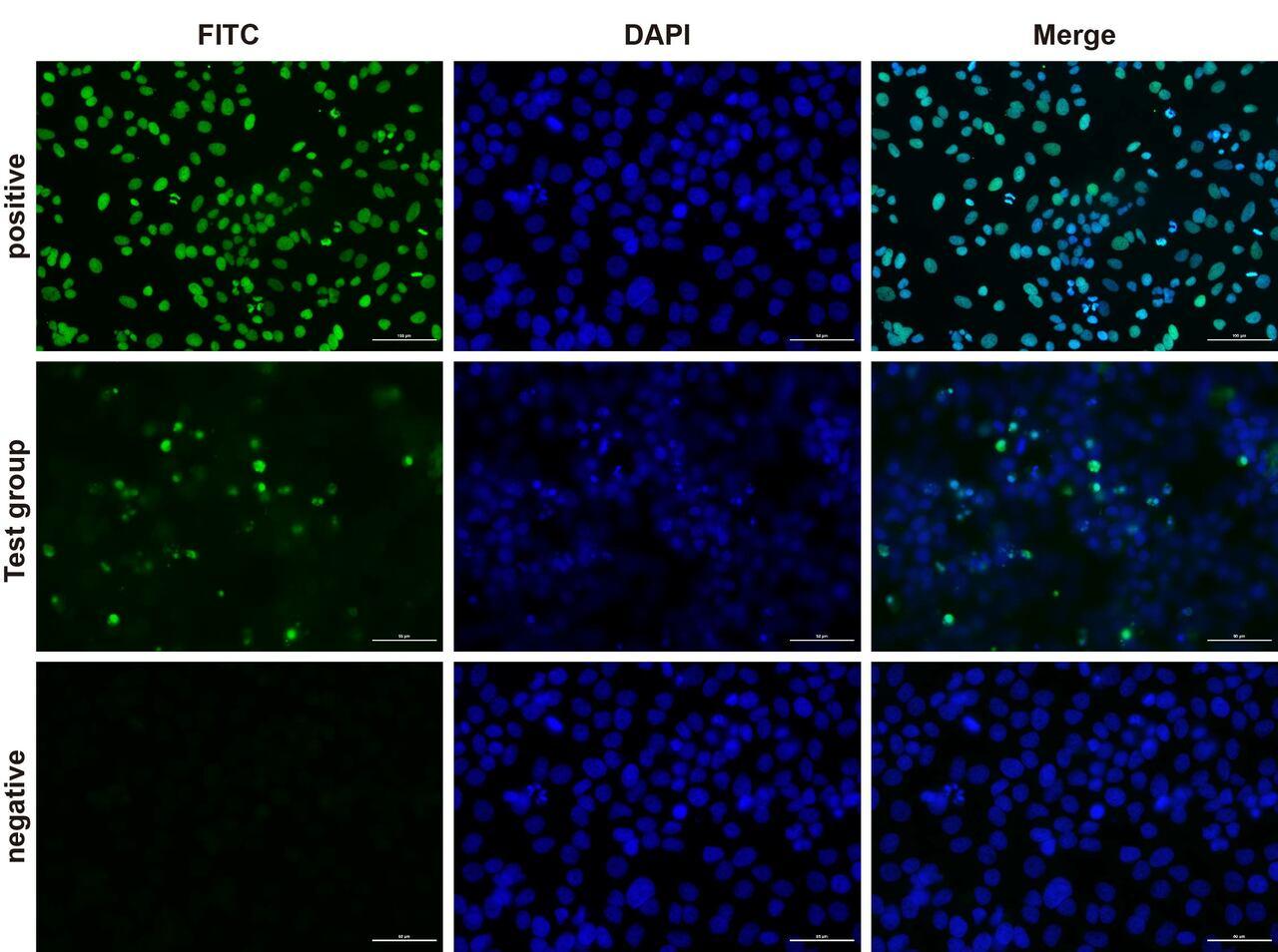 | Fluorescence microscope analysis of camptothecin-induced apoptosis of Hela cells. |


