Description
D-Lactic Acid/Lactate Colorimetric Assay Kit
The D-Lactic Acid/Lactate Colorimetric Assay Kit (MAES0015) quantifies D-lactic acid in serum, plasma, animal tissue and saliva samples on a microplate reader. D-lactate dehydrogenase converts D-lactic acid and NAD+ to pyruvic acid and NADH, and the reaction produces a purple compound measured at 530 nm. Researchers use the kit to study D-lactate levels in glycolysis and bacterial metabolism contexts.
D-lactic acid (D-LA), also called D-lactate, is the D-enantiomer of lactic acid and the target analyte of this kit. D-lactate dehydrogenase (D-LDH) catalyzes its reaction with NAD+, which converts D-lactic acid to pyruvic acid and NAD+ to NADH. The kit is intended to measure D-lactic acid content in tissue, serum, plasma, and saliva samples, and its listed research area is glycolysis and lipid metabolism.
Researchers use this kit to quantify D-lactic acid across the sample types listed for the assay, within the glycolysis and lipid metabolism research area. The supplied materials do not name specific disease models or physiological conditions, so the study context should be defined by the investigator. The kit is labeled for research use only and must not be used for clinical diagnosis.
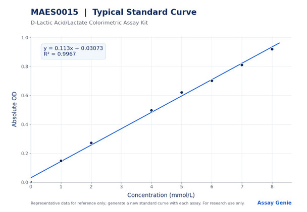
The assay uses NAD+ as the hydrogen (H+) acceptor. During the enzymatic reaction, NBT is reduced to a purple compound. In the procedure, 5 µL of standard or sample is combined with 100 µL of enzyme working solution and 20 µL of chromogenic agent, then incubated at 37°C for 10 min. After addition of 180 µL stop solution and mixing, absorbance is read at 530 nm. Concentrations are calculated from a standard curve built from an 8-point series of 0 to 8 mmol/L, prepared fresh for each assay.
Serum and plasma are assayed directly, and tissue and saliva require homogenization or clarification by centrifugation before testing. Severe hemolysis or jaundice may raise the OD value, so these conditions should be considered when interpreting results. Bubbles should be avoided when adding liquids to the microplate. If results are normalized to tissue protein, the protein concentration of each supernatant must be determined separately.
Reported analytical sensitivity is 0.06 mmol/L, with a detection range of 0.06 to 8.0 mmol/L. The product page lists intra-assay CV of 3.8% and inter-assay CV of 7.7%, and the manual reports intra-assay CV of 3.6 to 4.2% and inter-assay CV of 7.4 to 8.2% for human serum samples. Recovery across low, middle, and high concentration samples averaged 99%, with individual levels ranging from 98% to 101%.
| Product name: | D-Lactic Acid/Lactate Colorimetric Assay Kit |
| SKU: | MAES0015 |
| Size: | 96 Assays |
| Sample types: | Serum, plasma, animal tissue, saliva |
| Detection method: | Colorimetric |
| Instrument: | Microplate reader (520-550 nm; 530 nm optimum) |
| Sample volume: | 5 µL |
| Assay time: | 20 min |
| Sensitivity: | 0.06 mmol/L |
| Detection range: | 0.06-8.0 mmol/L |
| Research area: | Glycolysis & Carbohydrates |
| Sensitivity: | 0.06 mmol/L (two standard deviations above the mean OD of the zero standard, 20 replicates) |
| Detection range: | 0.06-8.0 mmol/L |
| Intra-assay CV: | 3.6-4.2% (three human serum samples, 20 replicates each) |
| Inter-assay CV: | 7.4-8.2% (three human serum samples, 20 assays in duplicate, three operators) |
| Recovery: | 98-101% (mean 99%; 6 replicates per level) |
- Colorimetric endpoint read at OD 530 nm (microplate reader, 520-550 nm range)
- Detection range 0.06-8.0 mmol/L; sensitivity 0.06 mmol/L
- Sample volume 5 µL; assay time 20 min
- Validated sample types: serum, plasma, animal tissue and saliva
- Supplied as 96 Assays with standard solution, buffer, enzyme, chromogenic agent and stop solution
Using NAD+ as the H+ acceptor, D-lactate dehydrogenase (D-LDH) catalyzes the reaction of D-lactic acid and NAD+ to generate pyruvic acid and NADH, respectively. During the reaction, NBT is reduced to a purple compound. The OD value is measured at 530 nm, and the D-lactic acid concentration is calculated from the standard curve.
- Quantifying D-lactic acid in serum and plasma from research animals and human samples
- Measuring D-lactate in animal tissue homogenates
- Studying D-lactate production linked to bacterial and gastrointestinal microbial metabolism
- Investigating glycolysis and carbohydrate metabolism pathways involving D-lactate
- Comparing D-lactate levels across experimental groups in saliva or tissue-derived samples
| Component | Quantity | Storage |
| Reagent 1: Buffer Solution | 12 mL x 1 vial | 2-8°C, 12 months |
| Reagent 2: Enzyme Stock Solution | 0.12 mL x 1 vial | 2-8°C, 12 months |
| Reagent 3: Chromogenic Agent | 1.2 mL x 2 vials | 2-8°C, 12 months; protect from light |
| Reagent 4: Stop Solution | 24 mL x 1 vial | 2-8°C, 12 months |
| Reagent 5: Standard Solution (10 mmol/L) | 2.0 mL x 1 vial | 2-8°C, 12 months |
| Microplate | 96 wells x 1 | No requirement |
| Plate sealer | 2 pieces | — |
| Sample layout sheet | 1 piece | — |
- Micropipettor
- Vortex mixer
- Incubator set to 37°C
- Centrifuge (including 10000 x g capability at 4°C)
- Microplate reader capable of 520-550 nm reading (530 nm optimum)
- Phosphate-buffered saline (0.01 mol/L, pH 7.4) for tissue preparation and sample dilution
- 0.9% NaCl (normal saline) as an alternative sample diluent
- Double-distilled water for standard dilutions
- Ice
- Dounce homogenizer (tissue samples)
- Separate protein concentration assay (required if results are normalized to protein)
Serum/plasma
- Detect serum or plasma directly.
- If samples are not detected on the same day, store serum or plasma at -80°C for up to one month.
- Recommended dilution factor is 1 for human plasma, human serum, rat serum, rat plasma, mouse serum, and rabbit serum.
- Use normal saline (0.9% NaCl) or PBS (0.01 M, pH 7.4) as the diluent. For other sample types, perform a pretest to confirm the dilution factor.
- Add 5 µL of sample per sample well.
Saliva
- Have the subject gargle with clear water.
- Collect the saliva 30 min later.
- Centrifuge at 10000 g for 10 min at 4°C.
- Take the supernatant and keep it on ice for detection.
- No dilution factor is listed for saliva; perform a pretest to confirm the dilution factor.
Tissue
- Harvest the amount of tissue needed for each assay (initial recommendation: 20 mg).
- Wash the tissue in cold PBS (0.01 M, pH 7.4).
- Homogenize 20 mg of tissue in 180 µL PBS (0.01 M, pH 7.4) with a Dounce homogenizer at 4°C.
- Centrifuge at 10000 × g for 10 min to remove insoluble material.
- Collect the supernatant and keep it on ice for detection.
- Determine the protein concentration of the supernatant separately.
- Recommended dilution factor is 2 to 3 for 10% rat kidney tissue homogenate and 1 for 10% mouse brain tissue homogenate.
- Use normal saline (0.9% NaCl) or PBS (0.01 M, pH 7.4) as the diluent. For other tissue types, perform a pretest to confirm the dilution factor.
Reagent preparation
- Equilibrate all reagents to room temperature before use.
- Keep the enzyme stock solution on ice until use.
- Prepare enzyme working solution fresh on the day of testing: mix 5 µL enzyme stock solution with 500 µL buffer solution per 5 µL of stock (for example, 505 µL total).
- Prepare a fresh set of standards for each assay and discard working dilutions after use.
- Dilute the 10 mmol/L standard with double-distilled water as follows: 0 mmol/L (0 µL standard + 200 µL water); 1 mmol/L (20 µL + 180 µL); 2 mmol/L (40 µL + 160 µL); 4 mmol/L (80 µL + 120 µL); 5 mmol/L (100 µL + 100 µL); 6 mmol/L (120 µL + 80 µL); 7 mmol/L (140 µL + 60 µL); 8 mmol/L (160 µL + 40 µL).
Sample preparation
- Serum and plasma: assay directly. If not assayed the same day, store at -80°C for up to one month.
- Saliva: have the subject gargle with clear water, collect saliva 30 min later, and centrifuge at 10000 g for 10 min at 4°C. Keep the supernatant on ice.
- Tissue: harvest the tissue needed for the assay (initial recommendation 20 mg) and wash in cold PBS (0.01 mol/L, pH 7.4).
- Tissue: homogenize 20 mg tissue in 180 µL PBS (0.01 mol/L, pH 7.4) with a Dounce homogenizer at 4°C.
- Tissue: centrifuge at 10000 x g for 10 min to remove insoluble material. Collect the supernatant and keep on ice.
- Dilute samples as needed: human plasma 1; human serum 1; rat serum 1; rat plasma 1; mouse serum 1; rabbit serum 1; 10% rat kidney tissue homogenate 2-3; 10% mouse brain tissue homogenate 1. Use normal saline or PBS (0.01 mol/L, pH 7.4) as diluent. Pretest other sample types to confirm dilution factor.
Assay procedure
- Add 5 µL of each standard to standard wells and 5 µL of each sample to sample wells.
- Add 100 µL enzyme working solution to each well.
- Add 20 µL chromogenic agent to each well.
- Mix thoroughly and incubate at 37°C for 10 min.
- Add 180 µL stop solution to each well.
- Mix thoroughly for 5 s and read OD values at 530 nm on a microplate reader.
Serum/plasma: D-LA content (mmol/L) = (ΔA530 - b) ÷ a × f Tissue: D-LA content (mmol/gprot) = (ΔA530 - b) ÷ a × f ÷ C_pr
- ΔA530 = OD530 Sample - OD530 Blank.
- a and b are the slope and intercept of the standard curve y = ax + b, where y is the absolute OD value of each standard (mean standard OD minus mean blank OD) and x is the standard concentration in mmol/L.
- f is the dilution factor of the sample before testing. For undiluted samples, f = 1.
- C_pr is the protein concentration of the sample in gprot/L, used only for tissue samples, which yields results in mmol/gprot.
Which sample types can be used with this kit?
The kit is intended for measuring D-lactic acid in tissue, serum, plasma, and saliva samples. The manual lists recommended dilution factors for human, rat, mouse, and rabbit serum or plasma and for 10% rat kidney and mouse brain tissue homogenates.
How should the kit and samples be stored?
Kit components should be stored at 2-8°C for 12 months, and the chromogenic agent must be kept protected from light. Serum or plasma that cannot be tested the same day can be stored at -80°C for up to one month.
What should I do if my results fall outside the detection range?
The detection range is 0.06 to 8.0 mmol/L. If the concentration is not within this range, take an extra dilution or concentration of the sample before testing.
What can interfere with the assay?
Severe hemolysis or jaundice may raise the OD value. Avoid forming bubbles when adding liquids to the microplate.
What instrument settings are required?
Read the plate with a microplate reader set between 520 and 550 nm, with an optimum wavelength of 530 nm.
How long does the assay take?
The listed assay time is 20 min. The protocol's incubation step is 10 min at 37°C.