The CD3D Monoclonal Antibody (CAB9770) is a high-quality antibody developed for reliable detection and analysis of target proteins. This polyclonal antibody, produced in rabbits, is highly specific to human CD3D and has been validated for use in various applications such as Western blot and immunofluorescence.CD3D is a critical component of the T cell receptor complex, essential for antigen recognition and signal transduction. Dysregulation of CD3D expression or function can lead to immune dysfunction and contribute to the development of autoimmune diseases or immune deficiencies.
This antibody is validated for use in WB, FC, ELISA, IF-P applications and has demonstrated reactivity against Human samples.
Product Name:
CD3D Monoclonal Antibody
SKU:
CAB9770
Size:
20μL, 100μL
Reactivity:
Human
Clone Number:
ARC1741
Conjugate:
Unconjugated
Immunogen:
Synthetic peptide. This information is considered to be commercially sensitive.
Recommended starting concentration is 1 μg/mL. Please optimize the concentration based on your specific assay requirements.
Synonyms:
T3D, IMD19, CD3DELTA, CD3-DELTA, CD3D
Positive Sample:
Jurkat
Cellular Localization:
Membrane, Single-Pass Type I Membrane Protein.
Calculated MW:
19kDa
Observed MW:
23kDa
The protein encoded by this gene is part of the T-cell receptor/CD3 complex (TCR/CD3 complex) and is involved in T-cell development and signal transduction. The encoded membrane protein represents the delta subunit of the CD3 complex, and along with four other CD3 subunits, binds either TCR alpha/beta or TCR gamma/delta to form the TCR/CD3 complex on the surface of T-cells. Defects in this gene are a cause of severe combined immunodeficiency autosomal recessive T-cell-negative/B-cell-positive/NK-cell-positive (SCIDBNK). Two transcript variants encoding different isoforms have been found for this gene. Other variants may also exist, but the full-length natures of their transcripts has yet to be defined.
Purification Method
Affinity purification
Gene ID
915
RRID
AB_2863777
Buffer Information
Store at -20℃. Avoid freeze / thaw cycles. Buffer: PBS containing 50% glycerol and 0.05% BSA, preserved with proclin300 or sodium azide, pH 7.3.
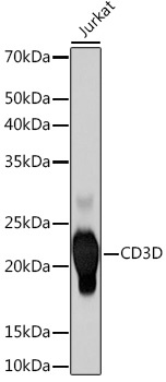
Western blot analysis of lysates from Jurkat cells, using CD3D Rabbit mAb (CAB9770) at 1:500 dilution. Secondary antibody: HRP-conjugated Goat anti-Rabbit IgG (H+L) (CABS014) at 1:10000 dilution. Lysates/proteins: 25μg per lane. Blocking buffer: 3% nonfat dry milk in TBST. Detection: ECL Basic Kit (AbGn00020). Exposure time: 60s.
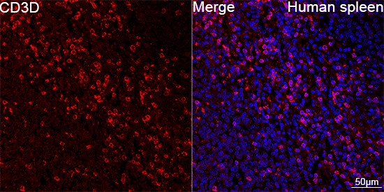
Confocal imaging of paraffin-embedded Human spleen tissue using CD3D Rabbit mAb (CAB9770, dilution 1:200) followed by a further incubation with Cy3 Goat Anti-Rabbit IgG (H+L) (CABS007, dilution 1:500) (Red). DAPI was used for nuclear staining (Blue). Objective: 40x. Perform high pressure antigen retrieval with 0.01 M citrate buffer (pH 6.0) prior to IF staining.


![Anti-CD3D [R03-2H9] Monoclonal Antibody (AGMB02837)](https://cdn11.bigcommerce.com/s-h68l9z2lnx/images/stencil/590x590/products/274126/679528/anti-cd3d-r03-2h9-monoclonal-antibody-agmb02837__00074.1773039248.jpg?c=2 "Anti-CD3D [R03-2H9] Monoclonal Antibody (AGMB02837)")
![Anti-CD3D [R09-6K4] Monoclonal Antibody (AGMB02041)](https://cdn11.bigcommerce.com/s-h68l9z2lnx/images/stencil/590x590/products/273330/676680/anti-cd3d-r09-6k4-monoclonal-antibody-agmb02041__74899.1773030248.jpg?c=2 "Anti-CD3D [R09-6K4] Monoclonal Antibody (AGMB02041)")
![Anti-CD3D [R02-7G-2] Monoclonal Antibody (AGMB03561)](https://cdn11.bigcommerce.com/s-h68l9z2lnx/images/stencil/590x590/products/274850/679383/anti-cd3d-r02-7g-2-monoclonal-antibody-agmb03561__99515.1773038774.jpg?c=2 "Anti-CD3D [R02-7G-2] Monoclonal Antibody (AGMB03561)")

