The CDH8 Antibody (CAB18691) is a high-quality antibody developed for reliable detection and analysis of target proteins. This antibody, raised in rabbits, is highly specific for human samples and has been validated for Western blot applications.CDH8, also known as Cadherin-8, is known to play a role in various cellular processes, including cell adhesion, migration, and signaling.
This antibody is validated for use in WB, IF/ICC, ELISA applications and has demonstrated reactivity against Mouse, Rat samples.
Product Name:
CDH8 Antibody
SKU:
CAB18691
Size:
20μL, 100μL
Reactivity:
Mouse, Rat
Immunogen:
Recombinant protein (or fragment).This information is considered to be commercially sensitive.
Tested Applications:
WBIF/ICCELISA
Recommended Dilution:
WB
1:500 - 1:2000
IF/ICC
1:50 - 1:200
ELISA
Recommended starting concentration is 1 μg/mL. Please optimize the concentration based on your specific assay requirements.
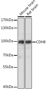
Positive Sample:
Mouse brain, Rat brain
Observed MW:
88kDa
This gene encodes a type II classical cadherin from the cadherin superfamily, integral membrane proteins that mediate calcium-dependent cell-cell adhesion. Mature cadherin proteins are composed of a large N-terminal extracellular domain, a single membrane-spanning domain, and a small, highly conserved C-terminal cytoplasmic domain. The extracellular domain consists of 5 subdomains, each containing a cadherin motif, and appears to determine the specificity of the protein's homophilic cell adhesion activity. Type II (atypical) cadherins are defined based on their lack of a HAV cell adhesion recognition sequence specific to type I cadherins. This particular cadherin is expressed in brain and is putatively involved in synaptic adhesion, axon outgrowth and guidance.
Purification Method
Affinity purification
Gene ID
1006
RRID
AB_2862425
Buffer Information
Store at -20℃. Avoid freeze / thaw cycles. Buffer: PBS with 0.01% thimerosal,50% glycerol,pH7.3.
Western blot analysis of various lysates using CDH8 Rabbit pAb (CAB18691) at 1:3000 dilution. Secondary antibody: HRP-conjugated Goat anti-Rabbit IgG (H+L) (CABS014) at 1:10000 dilution. Lysates/proteins: 25μg per lane. Blocking buffer: 3% nonfat dry milk in TBST. Detection: ECL Basic Kit (AbGn00020). Exposure time: 5s.
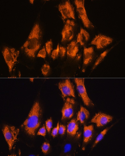
Immunofluorescence analysis of C6 cells using CDH8 Rabbit pAb (CAB18691) at dilution of 1:100. Secondary antibody: Cy3-conjugated Goat anti-Rabbit IgG (H+L) (CABS007) at 1:500 dilution. Blue: DAPI for nuclear staining.