The MMACHC Antibody (CAB10355) is a high-quality antibody developed for reliable detection and analysis of target proteins. This antibody, generated in rabbits, exhibits high reactivity to human samples and has been validated for use in Western blot applications. By specifically binding to the MMACHC protein, this antibody allows for precise detection and analysis in a variety of cell types, making it an essential component for investigations in biochemistry and molecular biology.MMACHC plays a crucial role in the intracellular processing of vitamin B12, a vital nutrient involved in various physiological processes, including DNA synthesis and neurological function. Dysregulation of MMACHC function has been linked to the development of disorders related to vitamin B12 deficiency, such as megaloblastic anemia and neurological complications.
This antibody is validated for use in WB, ELISA applications and has demonstrated reactivity against Human, Mouse, Rat samples.
Product Name:
MMACHC Antibody
SKU:
CAB10355
Size:
20μL, 100μL
Reactivity:
Human, Mouse, Rat
Conjugate:
Unconjugated
Immunogen:
Recombinant protein (or fragment).This information is considered to be commercially sensitive.
Recommended starting concentration is 1 μg/mL. Please optimize the concentration based on your specific assay requirements.
Synonyms:
cblC, MMACHC
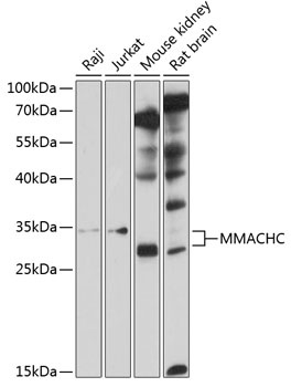
Positive Sample:
Raji, Jurkat, Mouse kidney, Rat brain
Cellular Localization:
Cytoplasm.
Calculated MW:
32kDa
Observed MW:
32kDa
The exact function of the protein encoded by this gene is not known, however, its C-terminal region shows similarity to TonB, a bacterial protein involved in energy transduction for cobalamin (vitamin B12) uptake. Hence, it is postulated that this protein may have a role in the binding and intracellular trafficking of cobalamin. Mutations in this gene are associated with methylmalonic aciduria and homocystinuria type cblC.
Purification Method
Affinity purification
Gene ID
25974
RRID
AB_2757900
Buffer Information
Store at -20℃. Avoid freeze / thaw cycles. Buffer: PBS containing 50% glycerol, preserved with proclin300 or sodium azide, pH 7.3.
Western blot analysis of various lysates using MMACHC Rabbit pAb (CAB10355) at 1:1000 dilution. Secondary antibody: HRP-conjugated Goat anti-Rabbit IgG (H+L) (CABS014) at 1:10000 dilution. Lysates/proteins: 25μg per lane. Blocking buffer: 3% nonfat dry milk in TBST. Detection: ECL Basic Kit (AbGn00020). Exposure time: 20s.