The NHLH1 Antibody (CAB15695) is a high-quality antibody developed for reliable detection and analysis of target proteins. This antibody, produced in rabbits, exhibits high reactivity with human samples and has been validated for use in Western blotting applications. By targeting the NHLH1 protein, this antibody allows for the detection and analysis of NHLH1 expression in various cell types, making it an ideal choice for studies in developmental biology and gene regulation.
This antibody is validated for use in WB, IF/ICC, IP, ELISA applications and has demonstrated reactivity against Human samples.
Product Name:
NHLH1 Antibody
SKU:
CAB15695
Size:
20μL, 100μL
Reactivity:
Human
Conjugate:
Unconjugated
Immunogen:
Recombinant protein (or fragment).This information is considered to be commercially sensitive.
0.5μg-4μg antibody for 200μg-400μg extracts of whole cells
ELISA
Recommended starting concentration is 1 μg/mL. Please optimize the concentration based on your specific assay requirements.
Synonyms:
HEN1, NSCL, NSCL1, bHLHa35, NHLH1
Positive Sample:
293T transfected with NHLH1
Cellular Localization:
Nucleus.
Calculated MW:
15kDa
Observed MW:
22kDa
The helix-loop-helix (HLH) proteins are a family of putative transcription factors, some of which have been shown to play an important role in growth and development of a wide variety of tissues and species. Four members of this family have been clearly implicated in tumorigenesis via their involvement in chromosomal translocations in lymphoid tumors: MYC (MIM 190080), LYL1 (MIM 151440), E2A (MIM 147141), and SCL (MIM 187040).
Purification Method
Affinity purification
Gene ID
4807
RRID
AB_2763106
Buffer Information
Store at -20℃. Avoid freeze / thaw cycles. Buffer: PBS containing 50% glycerol, preserved with proclin300 or sodium azide, pH 7.3.
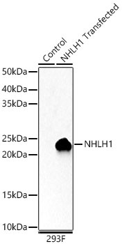
Western blot analysis of lysates from wild type (WT) and 293T cells transfected with NHLH1 using NHLH1 Rabbit pAb (CAB15695) at 1:3000 dilution. Secondary antibody: HRP-conjugated Goat anti-Rabbit IgG (H+L) (CABS014) at 1:10000 dilution. Lysates/proteins: 25μg per lane. Blocking buffer: 3% nonfat dry milk in TBST. Detection: ECL Basic Kit (AbGn00020). Exposure time: 30s.
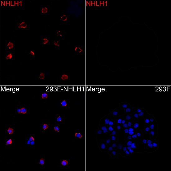
Immunofluorescence analysis of 293F cells transfected with NHLH1 using NHLH1 Rabbit pAb (CAB15695) at a dilution of 1:400 (40x lens). Secondary antibody: Cy3-conjugated Goat anti-Rabbit IgG (H+L)(CABS007) at 1:500 dilution. Blue: DAPI for nuclear staining.