The PGP9.5/UCHL1 Monoclonal Antibody (CAB19101) is a high-quality antibody developed for reliable detection and analysis of target proteins. This antibody, developed using rabbit monoclonal technology, shows high reactivity with human samples and is validated for use in various applications, including immunohistochemistry and ELISA.PGP9.5, also known as ubiquitin carboxyl-terminal hydrolase L1 (UCHL1), is a widely used neuronal marker due to its abundance in neurons and neuroendocrine cells.
This antibody is validated for use in WB, IHC-P, IF/ICC, ELISA, IF-P applications and has demonstrated reactivity against Human, Mouse, Rat, Monkey samples.
Product Name:
PGP9.5/UCHL1 Monoclonal Antibody
SKU:
CAB19101
Size:
20μL, 100μL
Reactivity:
Human, Mouse, Rat, Monkey
Clone Number:
ARC50371
Conjugate:
Unconjugated
Immunogen:
Recombinant protein (or fragment).This information is considered to be commercially sensitive.
The protein encoded by this gene belongs to the peptidase C12 family. This enzyme is a thiol protease that hydrolyzes a peptide bond at the C-terminal glycine of ubiquitin. This gene is specifically expressed in the neurons and in cells of the diffuse neuroendocrine system. Mutations in this gene may be associated with Parkinson disease.
Purification Method
Affinity purification
Gene ID
7345
RRID
AB_2862594
Buffer Information
Store at -20℃. Avoid freeze / thaw cycles. Buffer: PBS with 0.09% Sodium azide,0.05% BSA,50% glycerol,pH7.3.
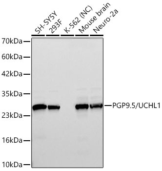
Western blot analysis of various lysates using PGP9.5/UCHL1 Rabbit mAb (CAB19101) at 1:120000 dilution incubated overnight at 4℃. Secondary antibody: HRP-conjugated Goat anti-Rabbit IgG (H+L) (CABS014) at 1:10000 dilution. Lysates/proteins: 25 μg per lane. Blocking buffer: 3% nonfat dry milk in TBST. Detection: ECL Basic Kit (AbGn00020). Negative control (NC): K-562 Exposure time: 30s.
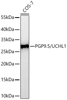
Western blot analysis of lysates from COS-7 cells using PGP9.5/UCHL1 Rabbit mAb (CAB19101) at 1:112000 dilution incubated overnight at 4℃. Secondary antibody: HRP-conjugated Goat anti-Rabbit IgG (H+L) (CABS014) at 1:10000 dilution. Lysates/proteins: 25 μg per lane. Blocking buffer: 3% nonfat dry milk in TBST. Detection: ECL Basic Kit (AbGn00020). Exposure time: 20s.
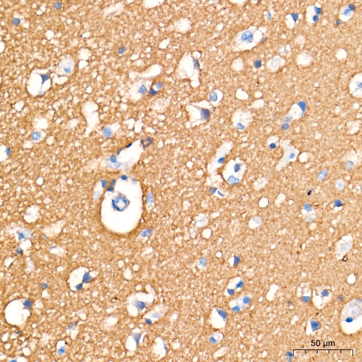
Immunohistochemistry analysis of paraffin-embedded Human brain tissue using PGP9.5/UCHL1 Rabbit mAb (CAB19101) at a dilution of 1:10000 (40x lens). High pressure antigen retrieval performed with 0.01M Tris-EDTA Buffer (pH 9.0) prior to IHC staining.
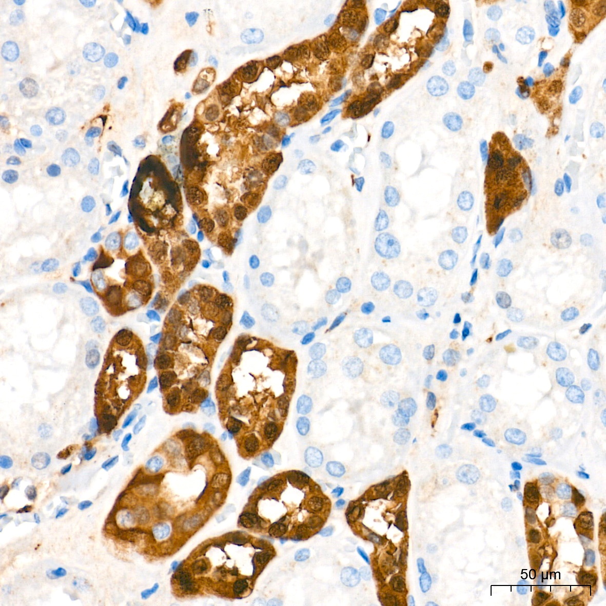
Immunohistochemistry analysis of paraffin-embedded Human kidney tissue using PGP9.5/UCHL1 Rabbit mAb (CAB19101) at a dilution of 1:10000 (40x lens). High pressure antigen retrieval performed with 0.01M Tris-EDTA Buffer (pH 9.0) prior to IHC staining.
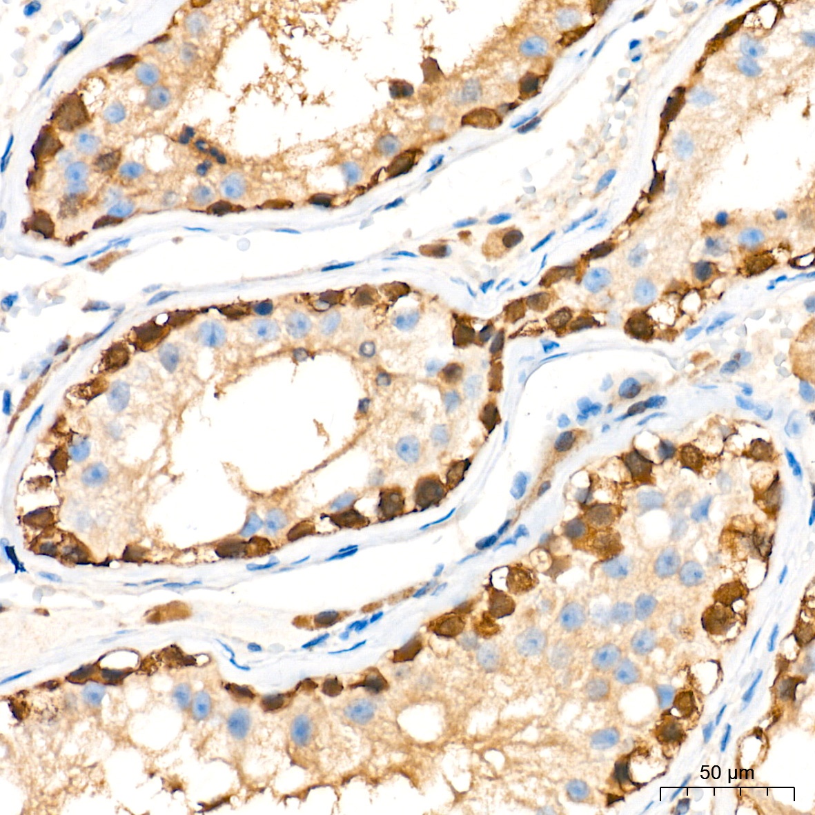
Immunohistochemistry analysis of paraffin-embedded Human testis tissue using PGP9.5/UCHL1 Rabbit mAb (CAB19101) at a dilution of 1:10000 (40x lens). High pressure antigen retrieval performed with 0.01M Tris-EDTA Buffer (pH 9.0) prior to IHC staining.
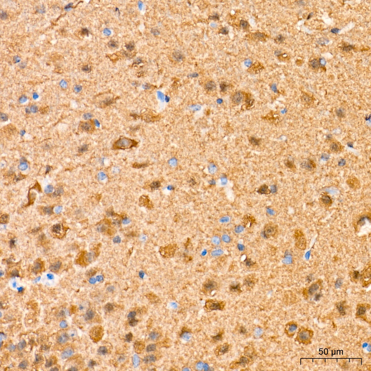
Immunohistochemistry analysis of paraffin-embedded Mouse brain tissue using PGP9.5/UCHL1 Rabbit mAb (CAB19101) at a dilution of 1:10000 (40x lens). High pressure antigen retrieval performed with 0.01M Tris-EDTA Buffer (pH 9.0) prior to IHC staining.
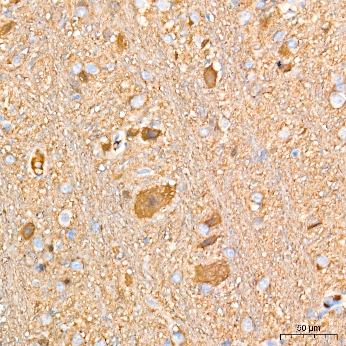
Immunohistochemistry analysis of paraffin-embedded Rat brain tissue using PGP9.5/UCHL1 Rabbit mAb (CAB19101) at a dilution of 1:10000 (40x lens). High pressure antigen retrieval performed with 0.01M Tris-EDTA Buffer (pH 9.0) prior to IHC staining.
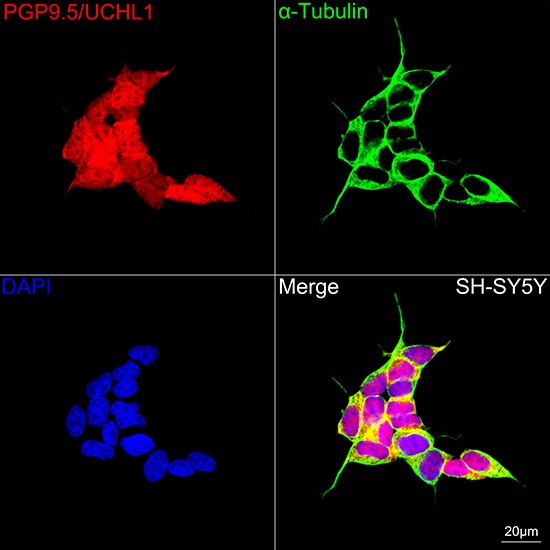
Confocal imaging of SH-SY5Y cells using PGP9.5/UCHL1 Rabbit mAb (CAB19101, dilution 1:500) followed by a further incubation with Cy3 Goat Anti-Rabbit IgG (H+L) (CABS007, dilution 1:500) (Red). The cells were counterstained with α-Tubulin Mouse mAb (AC012, dilution 1:400) followed by incubation with ABflo® 488-conjugated Goat Anti-Mouse IgG (H+L) Ab (CABS076, dilution 1:500) (Green). DAPI was used for nuclear staining (Blue). Objective: 100x.
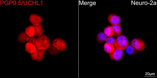
Confocal imaging of Neuro-2a cells using PGP9.5/UCHL1 Rabbit mAb (CAB19101, dilution 1:500) followed by a further incubation with Cy3 Goat Anti-Rabbit IgG (H+L) (CABS007, dilution 1:500) (Red). DAPI was used for nuclear staining (Blue). Objective: 100x.
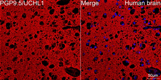
Confocal imaging of paraffin-embedded Human brain tissue using PGP9.5/UCHL1 Rabbit mAb (CAB19101, dilution 1:500) followed by a further incubation with Cy3 Goat Anti-Rabbit IgG (H+L) (CABS007, dilution 1:500) (Red). DAPI was used for nuclear staining (Blue). High pressure antigen retrieval performed with 0.01M Citrate Buffer (pH 6.0) prior to IF staining. Objective: 40x.
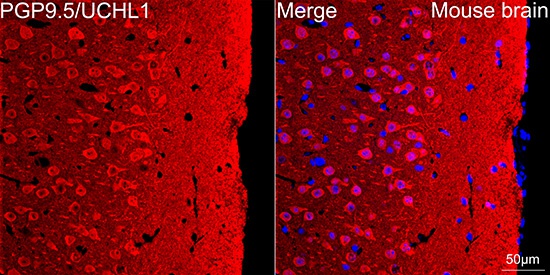
Confocal imaging of paraffin-embedded Mouse brain tissue using PGP9.5/UCHL1 Rabbit mAb (CAB19101, dilution 1:500) followed by a further incubation with Cy3 Goat Anti-Rabbit IgG (H+L) (CABS007, dilution 1:500) (Red). DAPI was used for nuclear staining (Blue). High pressure antigen retrieval performed with 0.01M Citrate Buffer (pH 6.0) prior to IF staining. Objective: 40x.


")
")
")