The Phospho-eIF2alpha-S51 Monoclonal Antibody (CABP0692) is a high-quality antibody developed for reliable detection and analysis of target proteins. This antibody, derived from rabbit monoclonal cells, exhibits high specificity and sensitivity for detecting phosphorylated eIF2alpha in human samples.Phosphorylation of eIF2alpha at serine 51 is a critical regulator of protein translation, controlling the synthesis of stress-response proteins in various cellular processes such as apoptosis, autophagy, and unfolded protein response. Dysregulation of eIF2alpha phosphorylation has been implicated in a range of diseases, including neurodegenerative disorders, viral infections, and cancer.
This antibody is validated for use in WB, ELISA applications and has demonstrated reactivity against Human, Mouse, Rat samples.
Product Name:
Phospho-eIF2alpha-S51 Monoclonal Antibody
SKU:
CABP0692
Size:
20μL, 100μL
Reactivity:
Human, Mouse, Rat
Clone Number:
ARC0130
Conjugate:
Unconjugated
Immunogen:
Synthetic peptide. This information is considered to be commercially sensitive.
Sequence:
Email for sequence
Tested Applications:
WBELISA
Recommended Dilution:
WB
1:2000 - 1:10000
ELISA
Recommended starting concentration is 1 μg/mL. Please optimize the concentration based on your specific assay requirements.
HeLa treated with Calyculin A, NIH/3T3 treated with Calyculin A, C6 treated with CA
Cellular Localization:
Cytoplasmic Granule.
Calculated MW:
36kDa
Observed MW:
36kDa
The translation initiation factor EIF2 catalyzes the first regulated step of protein synthesis initiation, promoting the binding of the initiator tRNA to 40S ribosomal subunits. Binding occurs as a ternary complex of methionyl-tRNA, EIF2, and GTP. EIF2 is composed of 3 nonidentical subunits, the 36-kD EIF2-alpha subunit (EIF2S1), the 38-kD EIF2-beta subunit (EIF2S2; MIM 603908), and the 52-kD EIF2-gamma subunit (EIF2S3; MIM 300161). The rate of formation of the ternary complex is modulated by the phosphorylation state of EIF2-alpha (Ernst et al., 1987 [PubMed 2948954]).
Purification Method
Affinity purification
Gene ID
1965
RRID
AB_2863809
Buffer Information
Store at -20℃. Avoid freeze / thaw cycles. Buffer: PBS with 0.09% sodium azide,0.05% BSA,50% glycerol,pH7.3.
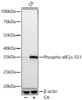
Western blot analysis of lysates from C6 cells using Phospho-eIF2α-S51 Rabbit mAb (CABP0692) at 1:10000 dilution incubated overnight at 4℃. C6 cells were treated with CA (100 nM) at 37℃ for 30 minutes after serum-starvation overnight. Secondary antibody: HRP-conjugated Goat anti-Rabbit IgG (H+L) (CABS014) at 1:10000 dilution. Lysates/proteins: 30 μg per lane. Blocking buffer: 3% nonfat dry milk in TBST. Detection: ECL Basic Kit (AbGn00020). Exposure time: 30 s.
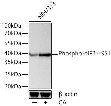
Western blot analysis of lysates from NIH/3T3 cells using Phospho-eIF2α-S51 Rabbit mAb (CABP0692) at 1:5000 dilution incubated at room temperature for 1.5 hours. NIH/3T3 cells were treated with CA (100 nM) at 37℃ for 30 minutes after serum-starvation overnight. Secondary antibody: HRP-conjugated Goat anti-Rabbit IgG (H+L) (CABS014) at 1:10000 dilution. Lysates/proteins: 30 μg per lane. Blocking buffer: 3 % nonfat dry milk in TBST. Detection: ECL Basic Kit (AbGn00020). Exposure time: 20 s.