The Rabbit IgG isotype control (CABC042) is a high-quality antibody developed for reliable detection and analysis of target proteins. This isotype control antibody, generated in rabbits, is highly specific for use in various research applications, including flow cytometry, immunohistochemistry, and immunoprecipitation.As a non-specific control, this Rabbit IgG Isotype Control ensures the accuracy and reliability of experimental results by binding nonspecifically to target molecules, allowing researchers to distinguish true antibody binding from background noise.
This antibody is validated for use in WB, IHC-P, IF/ICC, ChIP, FC, ELISA applications and has demonstrated reactivity against - samples.
Product Name:
Rabbit IgG isotype control
SKU:
CABC042
Size:
50μL, 100μL, 200μL
Clone Number:
ARC5105-03
Conjugate:
Unconjugated
Immunogen:
This information is considered to be commercially sensitive.
Tested Applications:
WBIHC-PIF/ICCChIPFCELISA
Recommended Dilution:
WB
1:5000 - 1:10000
IHC-P
1:50 - 1:200
IF/ICC
1:50 - 1:200
ChIP
3μg antibody for 10μg-15μg of Chromatin
ELISA
Recommended starting concentration is 1 μg/mL. Please optimize the concentration based on your specific assay requirements.
The protein encoded by this gene is a transcriptional regulator and tumor suppressor, serving as an activator of genes involved in both innate and acquired immune responses.
Purification Method
Protein A/G purification
Buffer Information
Store at -20℃. Avoid freeze / thaw cycles. Buffer: PBS with 0.09% sodium azid,0.05% BSA,50% glycerol,pH7.3.
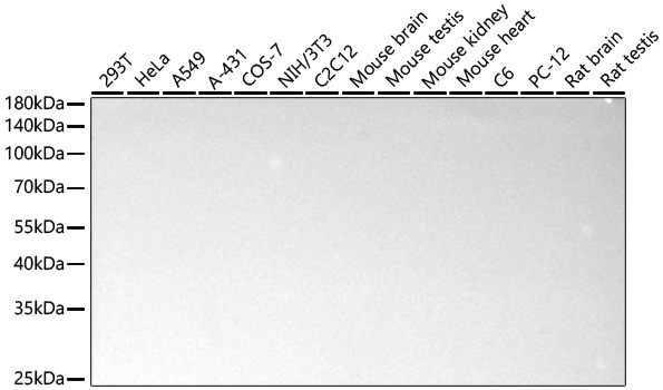
Western blot analysis of various lysates using Rabbit IgG isotype control (CABC042) at 1:10000 dilution incubated at room temperature for 1.5 hours. Secondary antibody: () at 1:10000 dilution. Lysates/proteins: 25 μg per lane. Blocking buffer: 3% nonfat dry milk in TBST. Detection: ECL Basic Kit (AbGn00020). Exposure time: 90s.
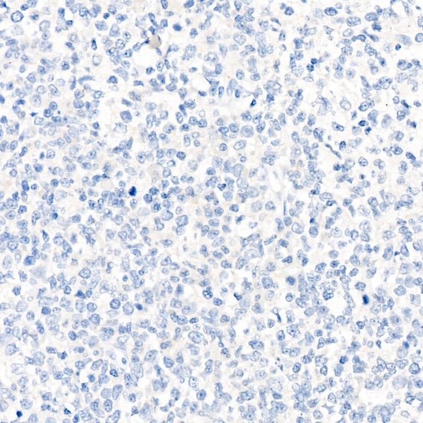
Immunohistochemistry analysis of paraffin-embedded Human tonsil using Rabbit IgG isotype control (CABC042) at dilution of 1:100 (40x lens). High pressure antigen retrieval performed with 0.01M Citrate buffer (pH 6.0) prior to IHC staining.
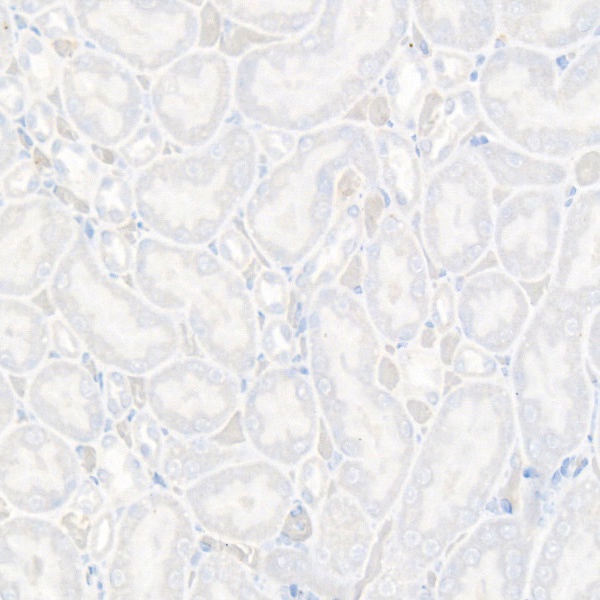
Immunohistochemistry analysis of paraffin-embedded Rat kidney using Rabbit IgG isotype control (CABC042) at dilution of 1:100 (40x lens). High pressure antigen retrieval performed with 0.01M Citrate buffer (pH 6.0) prior to IHC staining.
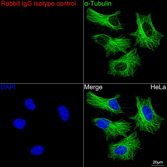
Confocal imaging of HeLa cells using Rabbit IgG isotype control (CABC042, dilution 1:200) followed by a further incubation with Cy3 Goat Anti-Rabbit IgG (H+L) (CABS007, dilution 1:500) (Red). The cells were counterstained with α-Tubulin Mouse mAb (AC012, dilution 1:400) followed by incubation with ABflo® 488-conjugated Goat Anti-Mouse IgG (H+L) Ab (CABS076, dilution 1:500) (Green). DAPI was used for nuclear staining (Blue). Objective: 100x.
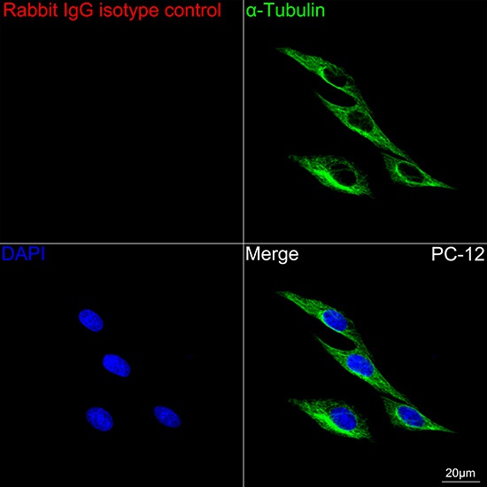
Confocal imaging of PC-12 cells using Rabbit IgG isotype control (CABC042, dilution 1:200) followed by a further incubation with Cy3 Goat Anti-Rabbit IgG (H+L) (CABS007, dilution 1:500) (Red). The cells were counterstained with α-Tubulin Mouse mAb (AC012, dilution 1:400) followed by incubation with ABflo® 488-conjugated Goat Anti-Mouse IgG (H+L) Ab (CABS076, dilution 1:500) (Green). DAPI was used for nuclear staining (Blue). Objective: 100x.
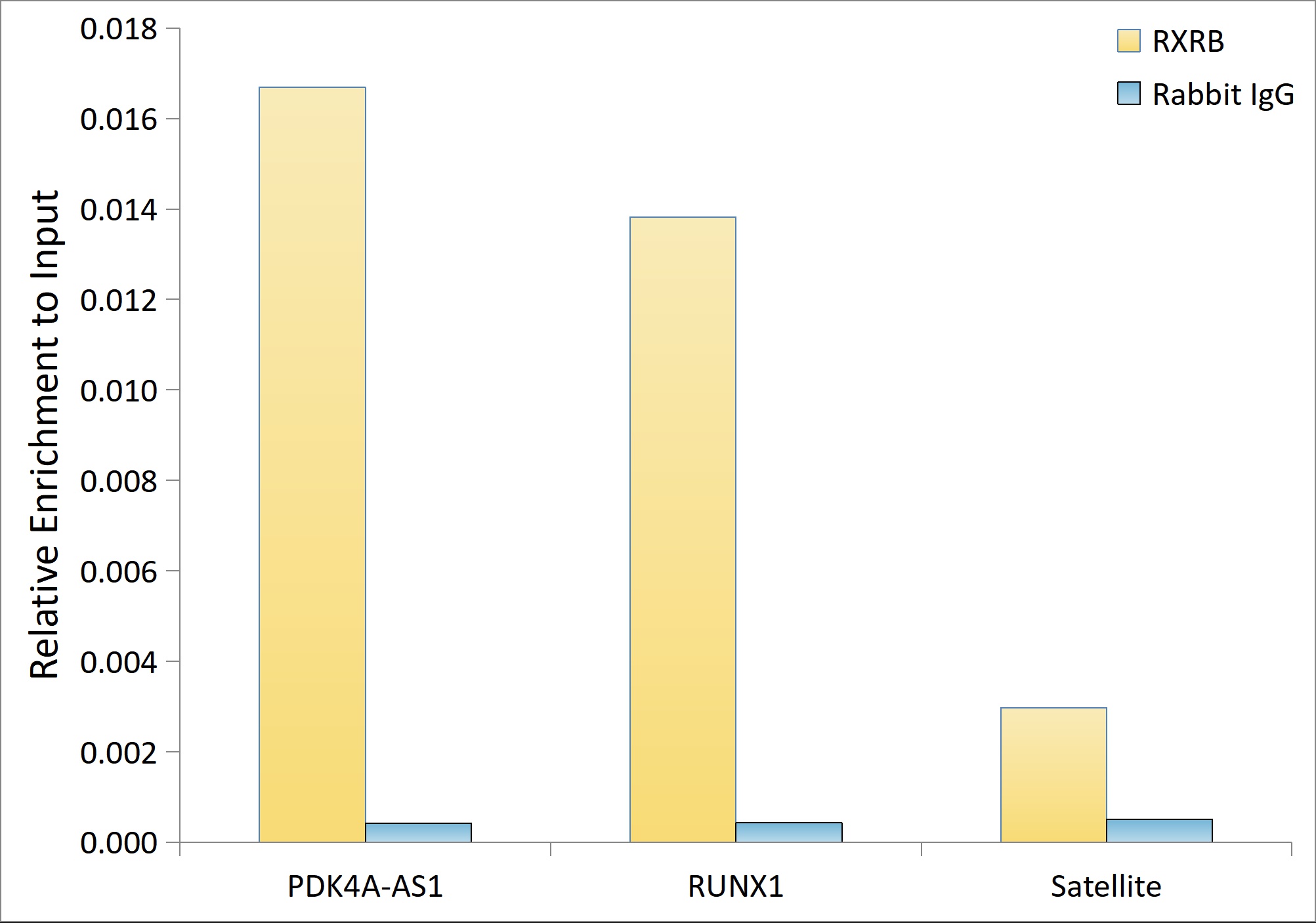
Chromatin immunoprecipitation was performed with 15 μg of cross-linked chromatin from HeLa cells transfected with a RXRB expression vector containing a single C-terminal flag-Tag, using 3 μg of Rabbit IgG isotype control (CABC042) and RXRB Rabbit mAb (CAB25648). The enrichment of immunoprecipitated DNA at different genomic loci was examined by quantitative PCR. The histogram compares the ratio of the immunoprecipitated DNA to the input at given loci.
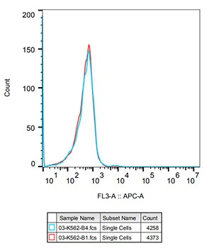
Flow cytometry: K562 cells were stained with Rabbit IgG isotype control(CABC042, 10 μg/mL, blue line) followed by goat anti-Rabbit pAb APC(1:600 dilution) staining. Non-fluorescently stained K562 cells was used as blank control (red line).
")
")
")
![FITC Armenian Hamster IgG Isotype Control [PIP] (AGEL2700)](https://cdn11.bigcommerce.com/s-h68l9z2lnx/images/stencil/590x590/products/229351/602226/fitc-armenian-hamster-igg-isotype-control-pip-agel2700__79473.1706284024.jpg?c=2 "FITC Armenian Hamster IgG Isotype Control [PIP] (AGEL2700)")
![FITC Armenian Hamster IgG Isotype Control [PIP] (AGEL2689)](https://cdn11.bigcommerce.com/s-h68l9z2lnx/images/stencil/590x590/products/229344/602303/fitc-armenian-hamster-igg-isotype-control-pip-agel2689__67507.1706284268.jpg?c=2 "FITC Armenian Hamster IgG Isotype Control [PIP] (AGEL2689)")
![APC Armenian Hamster IgG Isotype Control [PIP] (AGEL2702)](https://cdn11.bigcommerce.com/s-h68l9z2lnx/images/stencil/590x590/products/229353/602558/apc-armenian-hamster-igg-isotype-control-pip-agel2702__41172.1706285101.jpg?c=2 "APC Armenian Hamster IgG Isotype Control [PIP] (AGEL2702)")
")