The SP1 Antibody (CAB14662) is a high-quality antibody developed for reliable detection and analysis of target proteins. This antibody, generated in rabbits, is highly specific for SP1 and has been validated for use in immunohistochemistry and western blotting applications.SP1 is known for its role in transcriptional regulation, controlling the expression of genes involved in cell growth, differentiation, and apoptosis. Dysregulation of SP1 has been implicated in the development of cancer, inflammation, and other diseases, making it a promising target for therapeutic intervention.
This antibody is validated for use in WB, IHC-P, IF/ICC, ELISA applications and has demonstrated reactivity against Human, Mouse, Rat samples.
Product Name:
SP1 Antibody
SKU:
CAB14662
Size:
20μL, 100μL
Reactivity:
Human, Mouse, Rat
Conjugate:
Unconjugated
Immunogen:
Recombinant protein (or fragment).This information is considered to be commercially sensitive.
Recommended starting concentration is 1 μg/mL. Please optimize the concentration based on your specific assay requirements.
Synonyms:
SP1
Positive Sample:
Mouse heart
Cellular Localization:
Cytoplasm, Nucleus.
Calculated MW:
81kDa
Observed MW:
90kDa
The protein encoded by this gene is a zinc finger transcription factor that binds to GC-rich motifs of many promoters. The encoded protein is involved in many cellular processes, including cell differentiation, cell growth, apoptosis, immune responses, response to DNA damage, and chromatin remodeling. Post-translational modifications such as phosphorylation, acetylation, glycosylation, and proteolytic processing significantly affect the activity of this protein, which can be an activator or a repressor. Three transcript variants encoding different isoforms have been found for this gene.
Purification Method
Affinity purification
Gene ID
6667
RRID
AB_2761538
Buffer Information
Store at -20℃. Avoid freeze / thaw cycles. Buffer: PBS containing 50% glycerol, preserved with proclin300 or sodium azide, pH 7.3.
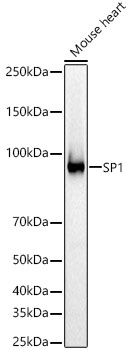
Western blot analysis of lysates from Mouse heart, using SP1 Rabbit pAb (CAB14662) at 1:600 dilution. Secondary antibody: HRP-conjugated Goat anti-Rabbit IgG (H+L) (CABS014) at 1:10000 dilution. Lysates/proteins: 25μg per lane. Blocking buffer: 3% nonfat dry milk in TBST. Detection: ECL Basic Kit (AbGn00020). Exposure time: 30s.
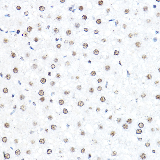
Immunohistochemistry analysis of paraffin-embedded Rat liver using SP1 Rabbit pAb (CAB14662) at dilution of 1:50 (40x lens). High pressure antigen retrieval performed with 0.01M Citrate buffer (pH 6.0) prior to IHC staining.
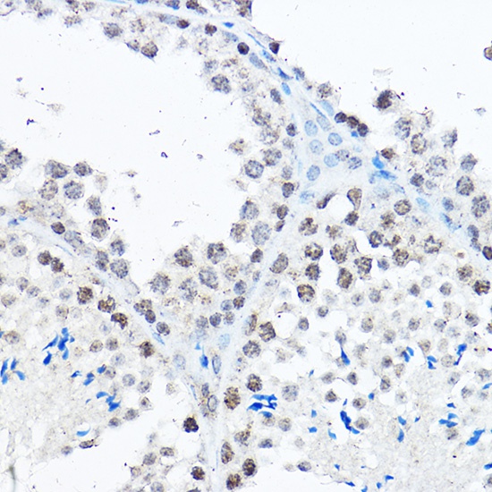
Immunohistochemistry analysis of paraffin-embedded Mouse testis using SP1 Rabbit pAb (CAB14662) at dilution of 1:50 (40x lens). High pressure antigen retrieval performed with 0.01M Citrate buffer (pH 6.0) prior to IHC staining.
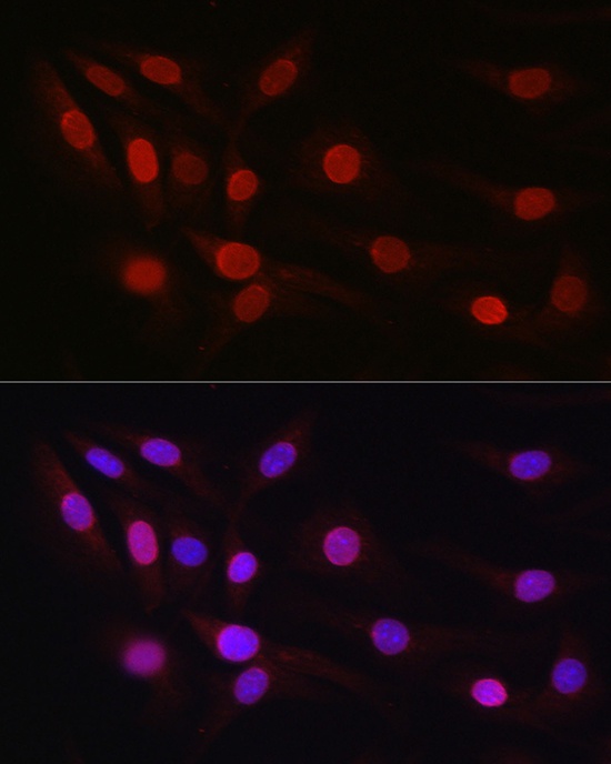
Immunofluorescence analysis of NIH/3T3 cells using SP1 Rabbit pAb (CAB14662) at dilution of 1:50 (40x lens). Secondary antibody: Cy3-conjugated Goat anti-Rabbit IgG (H+L) (CABS007) at 1:500 dilution. Blue: DAPI for nuclear staining.