The F4/80 Antibody (CAB18637) is a high-quality antibody developed for reliable detection and analysis of target proteins. Raised in rabbits, this antibody is highly specific for human samples and is validated for use in Western blot applications. By binding to the ADGRE1 protein, this antibody allows for precise detection and analysis in various cell types, making it well-suited for studies in immunology and cancer research.ADGRE1, also known as the adhesion G protein-coupled receptor E1, is known to play a crucial role in immune response modulation and cell adhesion processes.
This antibody is validated for use in WB, IF/ICC, ELISA applications and has demonstrated reactivity against Human, Mouse samples.
Product Name:
F4/80 Antibody
SKU:
CAB18637
Size:
20μL, 100μL
Reactivity:
Human, Mouse
Immunogen:
Recombinant protein (or fragment).This information is considered to be commercially sensitive.
External Side Of Plasma Membrane, Plasma Membrane.
Calculated MW:
102kDa
Observed MW:
65-250kDa
Predicted to enable G protein-coupled receptor activity. Predicted to be involved in adenylate cyclase-activating G protein-coupled receptor signaling pathway. Predicted to act upstream of or within G protein-coupled receptor signaling pathway and adaptive immune response. Located in external side of plasma membrane. Is expressed in several structures, including cardiovascular system; central nervous system; genitourinary system; hemolymphoid system; and intestine. Orthologous to human ADGRE1 (adhesion G protein-coupled receptor E1).
Purification Method
Affinity purification
Gene ID
13733
RRID
AB_2862375
Buffer Information
Store at -20℃. Avoid freeze / thaw cycles. Buffer: PBS containing 50% glycerol, preserved with proclin300 or sodium azide, pH 7.3.
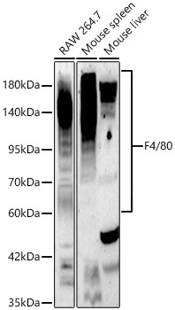
Western blot analysis of various lysates using F4/80 Rabbit pAb (CAB18637) at 1:2000 dilution. Secondary antibody: HRP-conjugated Goat anti-Rabbit IgG (H+L) (CABS014) at 1:10000 dilution. Lysates / proteins: 25 μg per lane. Blocking buffer: 3 % nonfat dry milk in TBST. Detection: ECL Basic Kit (AbGn00020). Exposure time: 90s.
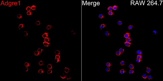
Immunofluorescence analysis of RAW264.7 cells using F4/80 Rabbit pAb (CAB18637) at dilution of 1:20 (40x lens). Secondary antibody: Cy3-conjugated Goat anti-Rabbit IgG (H+L) (CABS007) at 1:500 dilution. Blue: DAPI for nuclear staining.