The ADRM1 Antibody (CAB4481) is a high-quality antibody developed for reliable detection and analysis of target proteins. This antibody, produced in rabbits, exhibits high reactivity with human samples and has been validated for use in Western blot applications. By binding to the ADRM1 protein, this antibody enables the detection and analysis of ADRM1 in a variety of cell types, making it ideal for studies in molecular biology and protein degradation pathways.ADRM1, also known as the Proteasome 19S Regulatory Particle Non-ATPase Subunit 1, plays a key role in protein turnover by facilitating the recognition and processing of ubiquitinated substrates for degradation.
This antibody is validated for use in WB, IHC-P, IF/ICC, ELISA applications and has demonstrated reactivity against Human, Mouse, Rat samples.
Product Name:
ADRM1 Antibody
SKU:
CAB4481
Size:
20μL, 100μL
Reactivity:
Human, Mouse, Rat
Conjugate:
Unconjugated
Immunogen:
Recombinant protein (or fragment).This information is considered to be commercially sensitive.
Recommended starting concentration is 1 μg/mL. Please optimize the concentration based on your specific assay requirements.
Synonyms:
ARM1, ARM-1, GP110, PSMD16, ADRM1
Positive Sample:
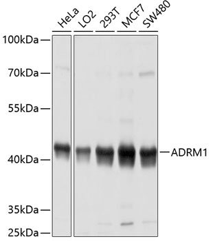
HeLa, LO2, 293T, MCF7, SW480
Cellular Localization:
Cytoplasm, Nucleus.
Calculated MW:
42kDa
Observed MW:
42kDa
This gene encodes a member of the adhesion regulating molecule 1 protein family. The encoded protein is a component of the proteasome where it acts as a ubiquitin receptor and recruits the deubiquitinating enzyme, ubiquitin carboxyl-terminal hydrolase L5. Increased levels of the encoded protein are associated with increased cell adhesion, which is likely an indirect effect of this intracellular protein. Dysregulation of this gene has been implicated in carcinogenesis. Alternative splicing results in multiple transcript variants.
Purification Method
Affinity purification
Gene ID
11047
RRID
AB_2765704
Buffer Information
Store at -20℃. Avoid freeze / thaw cycles. Buffer: PBS containing 50% glycerol, preserved with proclin300 or sodium azide, pH 7.3.
Western blot analysis of various lysates using ADAbGn1 Rabbit pAb (CAB4481) at 1:1000 dilution. Secondary antibody: HRP-conjugated Goat anti-Rabbit IgG (H+L) (CABS014) at 1:10000 dilution. Lysates/proteins: 25μg per lane. Blocking buffer: 3% nonfat dry milk in TBST. Detection: ECL Basic Kit (AbGn00020). Exposure time: 10s.
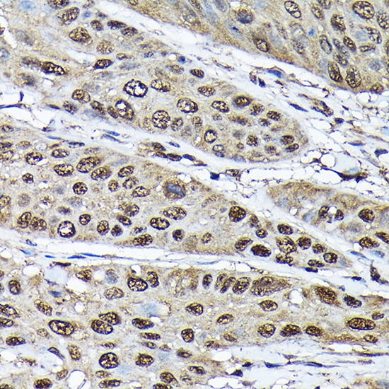
Immunohistochemistry analysis of paraffin-embedded Rat stomach using ADAbGn1 Rabbit pAb (CAB4481) at dilution of 1:100 (40x lens). High pressure antigen retrieval performed with 0.01M Citrate buffer (pH 6.0) prior to IHC staining.
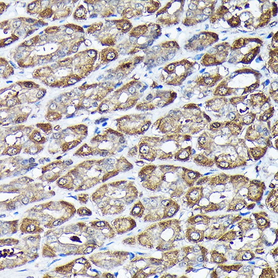
Immunohistochemistry analysis of paraffin-embedded Human esophageal cancer using ADAbGn1 Rabbit pAb (CAB4481) at dilution of 1:100 (40x lens). High pressure antigen retrieval performed with 0.01M Citrate buffer (pH 6.0) prior to IHC staining.
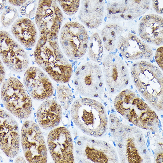
Immunohistochemistry analysis of paraffin-embedded Mouse kidney using ADAbGn1 Rabbit pAb (CAB4481) at dilution of 1:100 (40x lens). High pressure antigen retrieval performed with 0.01M Citrate buffer (pH 6.0) prior to IHC staining.