The AOX1 Antibody (CAB3586) is a high-quality antibody developed for reliable detection and analysis of target proteins. This antibody, generated in rabbits, exhibits high reactivity with human samples and has been validated for use in Western blot applications.Aldehyde oxidase plays a crucial role in drug metabolism and detoxification processes, making it a key target for pharmacological studies. By specifically binding to the aldehyde oxidase protein, this antibody enables researchers to detect and analyze its expression in different cell types and tissues, facilitating investigations into drug interactions, toxicity, and efficacy.
This antibody is validated for use in WB, IF/ICC, ELISA applications and has demonstrated reactivity against Human, Mouse, Rat samples.
Product Name:
AOX1 Antibody
SKU:
CAB3586
Size:
20μL, 100μL
Reactivity:
Human, Mouse, Rat
Conjugate:
Unconjugated
Immunogen:
Recombinant protein (or fragment).This information is considered to be commercially sensitive.
Recommended starting concentration is 1 μg/mL. Please optimize the concentration based on your specific assay requirements.
Synonyms:
AO, AOH1, AOX1
Positive Sample:
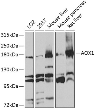
LO2, 293T, Mouse liver, Mouse pancreas, Rat liver
Cellular Localization:
Cytoplasm.
Calculated MW:
148kDa
Observed MW:
180kDa
Aldehyde oxidase produces hydrogen peroxide and, under certain conditions, can catalyze the formation of superoxide. Aldehyde oxidase is a candidate gene for amyotrophic lateral sclerosis.
Purification Method
Affinity purification
Gene ID
316
RRID
AB_2765167
Buffer Information
Store at -20℃. Avoid freeze / thaw cycles. Buffer: PBS containing 50% glycerol, preserved with proclin300 or sodium azide, pH 7.3.
Western blot analysis of various lysates using AOX1 Rabbit pAb (CAB3586) at 1:1000 dilution. Secondary antibody: HRP-conjugated Goat anti-Rabbit IgG (H+L) (CABS014) at 1:10000 dilution. Lysates/proteins: 25μg per lane. Blocking buffer: 3% nonfat dry milk in TBST. Detection: ECL Basic Kit (AbGn00020). Exposure time: 60s.
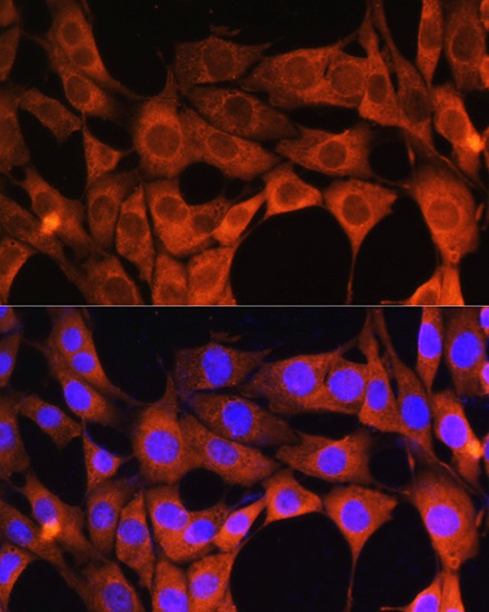
Immunofluorescence analysis of NIH/3T3 cells using AOX1 Rabbit pAb (CAB3586) at dilution of 1:100. Secondary antibody: Cy3-conjugated Goat anti-Rabbit IgG (H+L) (CABS007) at 1:500 dilution. Blue: DAPI for nuclear staining.
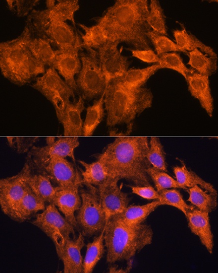
Immunofluorescence analysis of C6 cells using AOX1 Rabbit pAb (CAB3586) at dilution of 1:100. Secondary antibody: Cy3-conjugated Goat anti-Rabbit IgG (H+L) (CABS007) at 1:500 dilution. Blue: DAPI for nuclear staining.