The 5HT7 Receptor Monoclonal Antibody (CAB19706) is a high-quality antibody developed for reliable detection and analysis of target proteins. This antibody, produced through immunization of rabbits, is highly specific to human samples and has been validated for Western blot applications.The 5HT7 receptor is known to be involved in various physiological processes, including mood regulation, sleep-wake cycle, and cognitive function. Dysregulation of this receptor has been implicated in psychiatric disorders such as depression, anxiety, and schizophrenia.
This antibody is validated for use in WB, ELISA, IF-P applications and has demonstrated reactivity against Human, Mouse, Rat samples.
Product Name:
5HT7 Receptor Monoclonal Antibody
SKU:
CAB19706
Size:
20μL, 100μL
Reactivity:
Human, Mouse, Rat
Clone Number:
ARC2238
Conjugate:
Unconjugated
Immunogen:
Synthetic peptide. This information is considered to be commercially sensitive.
The neurotransmitter, serotonin, is thought to play a role in various cognitive and behavioral functions. The serotonin receptor encoded by this gene belongs to the superfamily of G protein-coupled receptors and the gene is a candidate locus for involvement in autistic disorder and other neuropsychiatric disorders. Three splice variants have been identified which encode proteins that differ in the length of their carboxy terminal ends.
Purification Method
Affinity purification
Gene ID
3363
Buffer Information
Store at -20℃. Avoid freeze / thaw cycles. Buffer: PBS containing 50% glycerol and 0.05% BSA, preserved with proclin300 or sodium azide, pH 7.3.
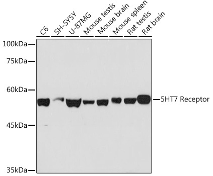
Western blot analysis of various lysates using 5HT7 Receptor Rabbit mAb (CAB19706) at 1:1000 dilution. Secondary antibody: HRP-conjugated Goat anti-Rabbit IgG (H+L) (CABS014) at 1:10000 dilution. Lysates/proteins: 25μg per lane. Blocking buffer: 3% nonfat dry milk in TBST. Detection: ECL Basic Kit (AbGn00020). Exposure time: 90s.
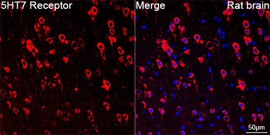
Confocal imaging of paraffin-embedded Rat brain using 5HT7 Receptor Rabbit mAb (CAB19706,dilution 1:100) followed by a further incubation with Cy3 Goat Anti-Rabbit IgG (H+L) (CABS007,dilution 1:500)(Red).DAPI was used for nuclear staining (Blue). Objective: 40x. Perform microwave antigen retrieval with 0.01 M citRate buffer (pH 6.0) prior to IF staining.