The CHD3 Monoclonal Antibody (CAB6118) is a high-quality antibody developed for reliable detection and analysis of target proteins. This antibody, produced in rabbits, demonstrates high specificity and sensitivity for human samples, making it suitable for use in Western blot applications.CHD3 is essential for maintaining chromatin structure and regulating gene expression, impacting various cellular processes such as cell differentiation, DNA repair, and development.
This antibody is validated for use in WB, IHC-P, ELISA, IF-P applications and has demonstrated reactivity against Human, Mouse, Rat samples.
Product Name:
CHD3 Monoclonal Antibody
SKU:
CAB6118
Size:
20μL, 100μL
Reactivity:
Human, Mouse, Rat
Clone Number:
ARC1975
Conjugate:
Unconjugated
Immunogen:
Synthetic peptide. This information is considered to be commercially sensitive.
This gene encodes a member of the CHD family of proteins which are characterized by the presence of chromo (chromatin organization modifier) domains and SNF2-related helicase/ATPase domains. This protein is one of the components of a histone deacetylase complex referred to as the Mi-2/NuRD complex which participates in the remodeling of chromatin by deacetylating histones. Chromatin remodeling is essential for many processes including transcription. Autoantibodies against this protein are found in a subset of patients with dermatomyositis. Three alternatively spliced transcripts encoding different isoforms have been described.
Purification Method
Affinity purification
Gene ID
1107
RRID
AB_2863523
Buffer Information
Store at -20℃. Avoid freeze / thaw cycles. Buffer: PBS containing 50% glycerol and 0.05% BSA, preserved with proclin300 or sodium azide, pH 7.3.
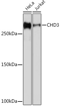
Western blot analysis of various lysates using CHD3 Rabbit mAb (CAB6118) at 1:1000 dilution. Secondary antibody: HRP-conjugated Goat anti-Rabbit IgG (H+L) (CABS014) at 1:10000 dilution. Lysates/proteins: 25μg per lane. Blocking buffer: 3% nonfat dry milk in TBST. Detection: ECL Basic Kit (AbGn00020). Exposure time: 1s.
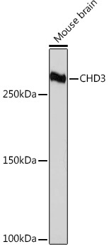
Western blot analysis of lysates from Mouse brain, using CHD3 Rabbit mAb (CAB6118) at 1:1000 dilution. Secondary antibody: HRP-conjugated Goat anti-Rabbit IgG (H+L) (CABS014) at 1:10000 dilution. Lysates/proteins: 25μg per lane. Blocking buffer: 3% nonfat dry milk in TBST. Detection: ECL Basic Kit (AbGn00020). Exposure time: 3min.
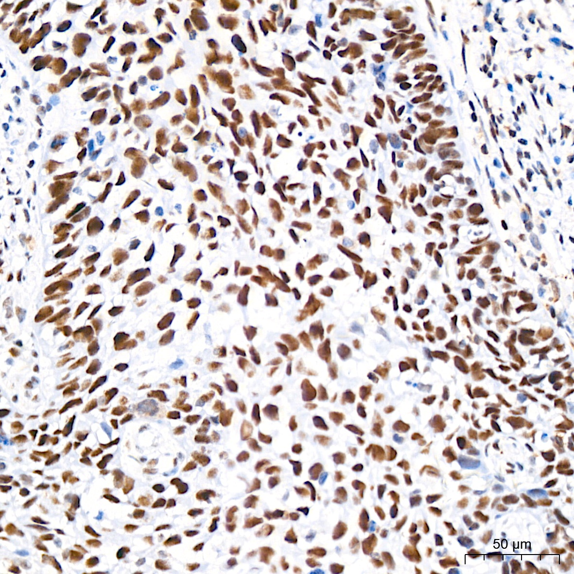
Immunohistochemistry analysis of paraffin-embedded Human cervix cancer tissue using CHD3 Rabbit mAb (CAB6118) at a dilution of 1:200 (40x lens). High pressure antigen retrieval performed with 0.01M Citrate buffer (pH 6.0) prior to IHC staining.
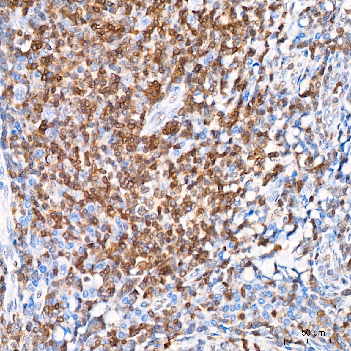
Immunohistochemistry analysis of paraffin-embedded Human tonsil tissue using CHD3 Rabbit mAb (CAB6118) at a dilution of 1:200 (40x lens). High pressure antigen retrieval performed with 0.01M Citrate buffer (pH 6.0) prior to IHC staining.
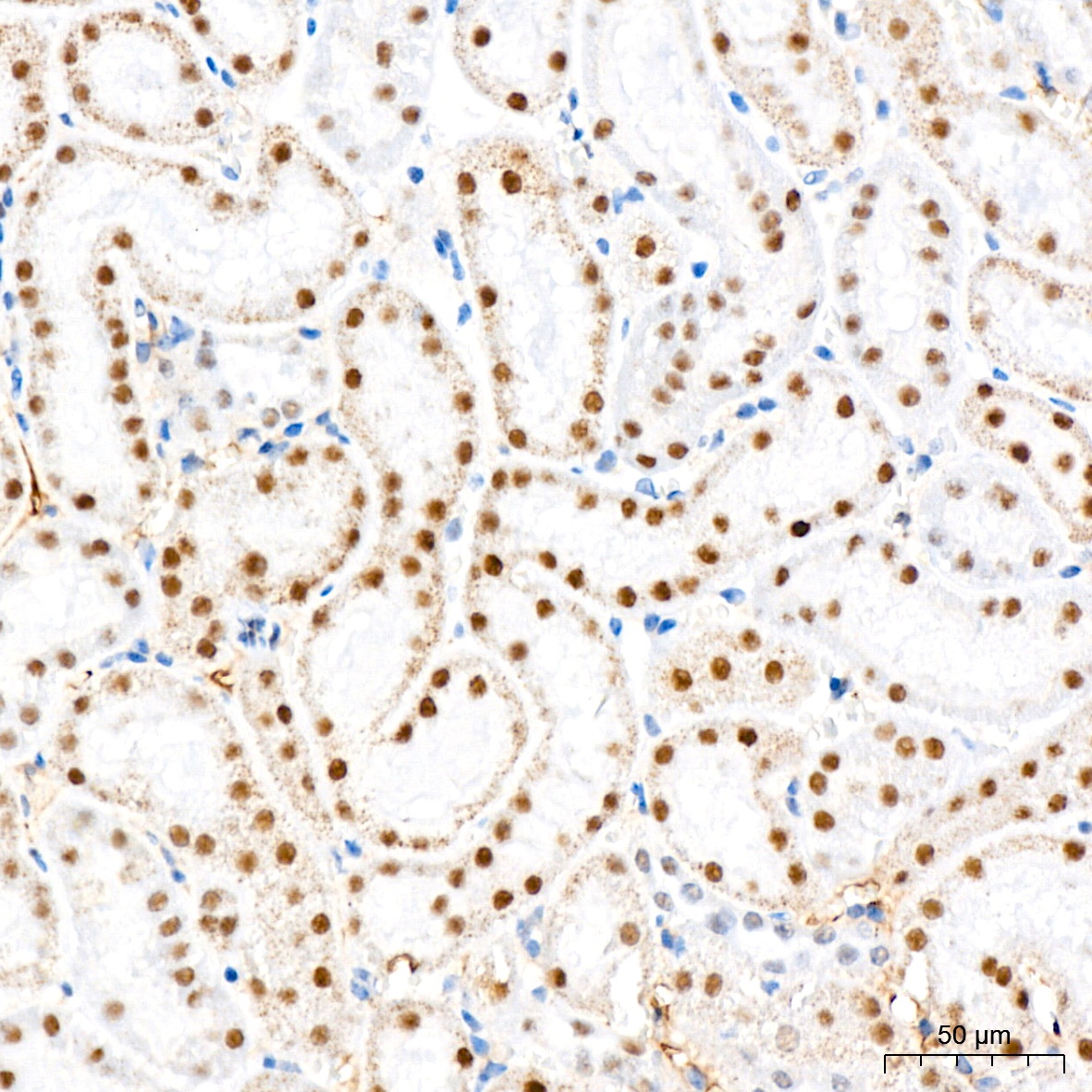
Immunohistochemistry analysis of paraffin-embedded Mouse kidney tissue using CHD3 Rabbit mAb (CAB6118) at a dilution of 1:200 (40x lens). High pressure antigen retrieval performed with 0.01M Citrate buffer (pH 6.0) prior to IHC staining.
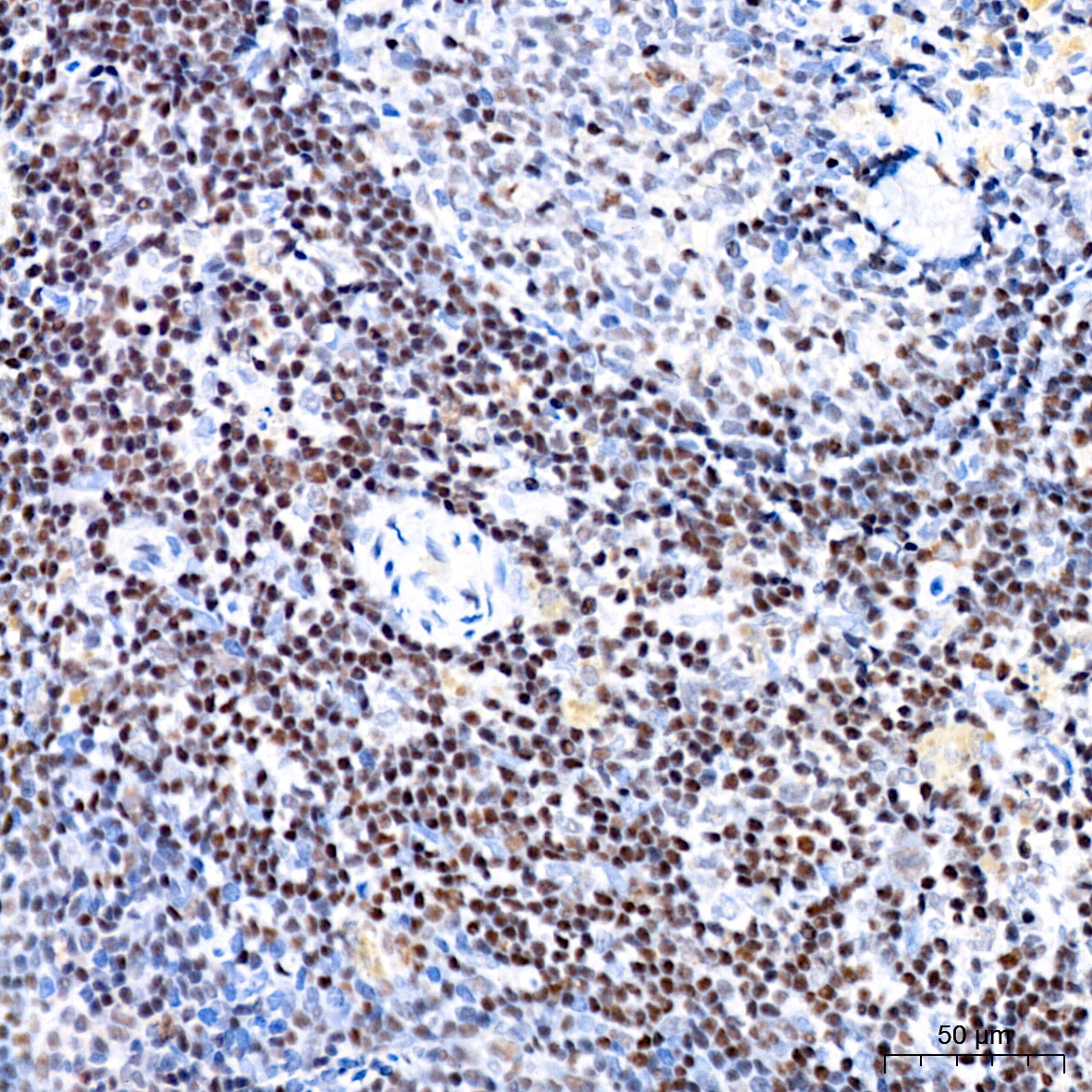
Immunohistochemistry analysis of paraffin-embedded Rat spleen tissue using CHD3 Rabbit mAb (CAB6118) at a dilution of 1:200 (40x lens). High pressure antigen retrieval performed with 0.01M Citrate buffer (pH 6.0) prior to IHC staining.
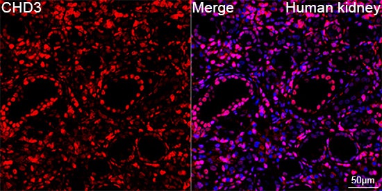
Confocal imaging of paraffin-embedded Human kidney using CHD3 Rabbit mAb (CAB6118,dilution 1:100) followed by a further incubation with Cy3 Goat Anti-Rabbit IgG (H+L) (CABS007,dilution 1:500)(Red).DAPI was used for nuclear staining (Blue). Objective: 40x. Perform high pressure antigen retrieval with 0.01 M citRate buffer (pH 6.0) prior to IF staining.
Confocal imaging of paraffin-embedded Human kidney using CHD3 Rabbit mAb (CAB6118,dilution 1:100) followed by a further incubation with Cy3 Goat Anti-Rabbit IgG (H+L) (CABS007,dilution 1:500)(Red).DAPI was used for nuclear staining (Blue). Objective: 40x. Perform high pressure antigen retrieval with 0.01 M citRate buffer (pH 6.0) prior to IF staining.


![Anti-CHD3 [R05-6G3] Monoclonal Antibody (AGMB00912)](https://cdn11.bigcommerce.com/s-h68l9z2lnx/images/stencil/590x590/products/272201/690704/anti-chd3-r05-6g3-monoclonal-antibody-agmb00912__76128.1774500683.jpg?c=2 "Anti-CHD3 [R05-6G3] Monoclonal Antibody (AGMB00912)")
![Anti-CHD3 [R03-4P-4] Monoclonal Antibody (AGMB03862)](https://cdn11.bigcommerce.com/s-h68l9z2lnx/images/stencil/590x590/products/275151/679858/anti-chd3-r03-4p-4-monoclonal-antibody-agmb03862__90330.1773040232.jpg?c=2 "Anti-CHD3 [R03-4P-4] Monoclonal Antibody (AGMB03862)")
![Anti-CHD3 (7F6) [7F6-G2-E5] Monoclonal Antibody (AGMB04245)](https://cdn11.bigcommerce.com/s-h68l9z2lnx/images/stencil/590x590/products/275534/679380/anti-chd3-7f6-7f6-g2-e5-monoclonal-antibody-agmb04245__22448.1773038771.jpg?c=2 "Anti-CHD3 (7F6) [7F6-G2-E5] Monoclonal Antibody (AGMB04245)")

