The HSP70/HSPA1 Antibody (CAB12948) is a high-quality antibody developed for reliable detection and analysis of target proteins. This antibody, produced in rabbits, exhibits high reactivity with human samples and is validated for use in Western blot applications. By binding specifically to Hsp70, researchers can accurately detect and analyze the protein in a variety of cell types.Hsp70 is known for its role in protecting cells from stress-induced damage and promoting cell survival. Research has shown that dysregulation of Hsp70 expression is linked to various diseases, including cancer, neurodegenerative disorders, and cardiovascular diseases.
This antibody is validated for use in WB, IHC-P, IF/ICC, ELISA applications and has demonstrated reactivity against Human, Mouse, Rat samples.
Product Name:
HSP70/HSPA1 Antibody
SKU:
CAB12948
Size:
20μL, 100μL
Reactivity:
Human, Mouse, Rat
Conjugate:
Unconjugated
Immunogen:
Synthetic peptide. This information is considered to be commercially sensitive.
Sequence:
KITI TNDK GRLS KEEI ERMV QEAE KYKA EDEV QRER VSAK NALE SYAF NMKS AVED EGLK GKIS EADK KKVL DKCQ EVIS WLDA NTLA EKDE FEHK RKEL E
Tested Applications:
WBIHC-PIF/ICCELISA
Recommended Dilution:
WB
1:1000 - 1:3000
IHC-P
1:50 - 1:200
IF/ICC
1:50 - 1:200
ELISA
Recommended starting concentration is 1 μg/mL. Please optimize the concentration based on your specific assay requirements.
This intronless gene encodes a 70kDa heat shock protein which is a member of the heat shock protein 70 family. In conjuction with other heat shock proteins, this protein stabilizes existing proteins against aggregation and mediates the folding of newly translated proteins in the cytosol and in organelles. It is also involved in the ubiquitin-proteasome pathway through interaction with the AU-rich element RNA-binding protein 1. The gene is located in the major histocompatibility complex class III region, in a cluster with two closely related genes which encode similar proteins.
Purification Method
Affinity purification
Gene ID
3303 3304
RRID
AB_2759794
Buffer Information
Store at -20℃. Avoid freeze / thaw cycles. Buffer: PBS containing 50% glycerol, preserved with proclin300 or sodium azide, pH 7.3.
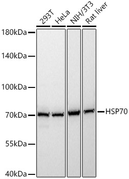
Western blot analysis of various lysates using HSP70 Rabbit pAb (CAB12948) at 1:10000 dilution incubated overnight at 4℃. Secondary antibody: HRP-conjugated Goat anti-Rabbit IgG (H+L) (CABS014) at 1:10000 dilution. Lysates/proteins: 25 μg per lane. Blocking buffer: 3% nonfat dry milk in TBST. Detection: ECL Basic Kit (AbGn00020). Exposure time: 1s.
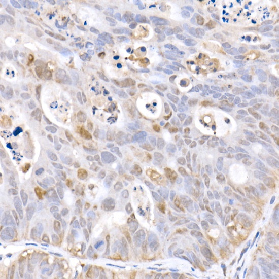
Immunohistochemistry analysis of paraffin-embedded Human colon carcinoma tissue using HSP70 Rabbit pAb (CAB12948) at a dilution of 1:100 (40x lens). High pressure antigen retrieval was performed with 0.01 M citrate buffer (pH 6.0) prior to IHC staining.
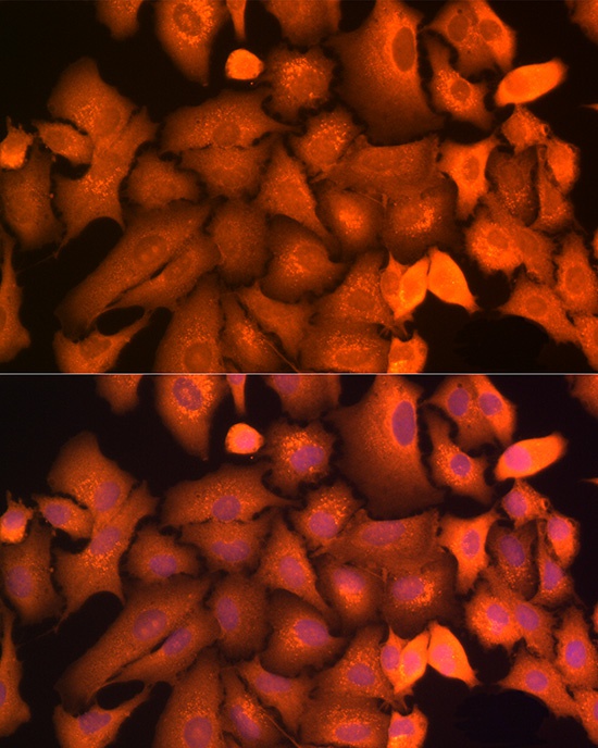
Immunofluorescence analysis of A-549 cells using HSP70 Rabbit pAb (CAB12948) at dilution of 1:100 (40x lens). Secondary antibody: Cy3-conjugated Goat anti-Rabbit IgG (H+L) (CABS007) at 1:500 dilution. Blue: DAPI for nuclear staining.
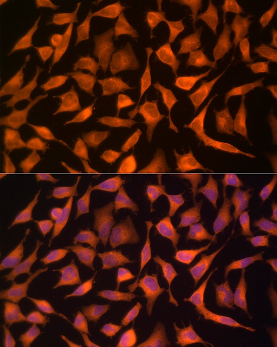
Immunofluorescence analysis of HeLa cells using HSP70 Rabbit pAb (CAB12948) at dilution of 1:100 (40x lens). Secondary antibody: Cy3-conjugated Goat anti-Rabbit IgG (H+L) (CABS007) at 1:500 dilution. Blue: DAPI for nuclear staining.
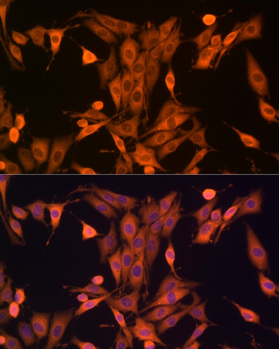
Immunofluorescence analysis of PC-12 cells using HSP70 Rabbit pAb (CAB12948) at dilution of 1:100 (40x lens). Secondary antibody: Cy3-conjugated Goat anti-Rabbit IgG (H+L) (CABS007) at 1:500 dilution. Blue: DAPI for nuclear staining.
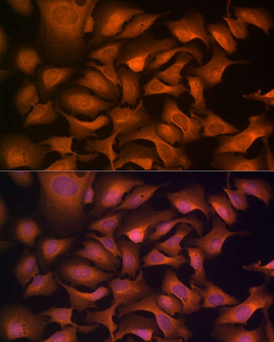
Immunofluorescence analysis of U2OS cells using HSP70 Rabbit pAb (CAB12948) at dilution of 1:100 (40x lens). Secondary antibody: Cy3-conjugated Goat anti-Rabbit IgG (H+L) (CABS007) at 1:500 dilution. Blue: DAPI for nuclear staining.


![Anti-Hsp70 [R04-7A2] Monoclonal Antibody (AGMB02176)](https://cdn11.bigcommerce.com/s-h68l9z2lnx/images/stencil/590x590/products/273465/677839/anti-hsp70-r04-7a2-monoclonal-antibody-agmb02176__26537.1773033869.jpg?c=2 "Anti-Hsp70 [R04-7A2] Monoclonal Antibody (AGMB02176)")
![Anti-Hsp70 [R07-6E9] Monoclonal Antibody (AGMB01699)](https://cdn11.bigcommerce.com/s-h68l9z2lnx/images/stencil/590x590/products/272988/678505/anti-hsp70-r07-6e9-monoclonal-antibody-agmb01699__78314.1773036011.jpg?c=2 "Anti-Hsp70 [R07-6E9] Monoclonal Antibody (AGMB01699)")
![Anti-Hsp70 [R01-5N-8] Monoclonal Antibody (AGMB03641)](https://cdn11.bigcommerce.com/s-h68l9z2lnx/images/stencil/590x590/products/274930/676531/anti-hsp70-r01-5n-8-monoclonal-antibody-agmb03641__62297.1773029771.jpg?c=2 "Anti-Hsp70 [R01-5N-8] Monoclonal Antibody (AGMB03641)")
![Anti-Hsp70 1A (2A11) [2A11-F3-G12] Monoclonal Antibody (AGMB04509)](https://cdn11.bigcommerce.com/s-h68l9z2lnx/images/stencil/590x590/products/275797/676871/anti-hsp70-1a-2a11-2a11-f3-g12-monoclonal-antibody-agmb04509__80799.1773030851.jpg?c=2 "Anti-Hsp70 1A (2A11) [2A11-F3-G12] Monoclonal Antibody (AGMB04509)")
![Anti-Hsp70 1A (2D4) [2D4-F7-C11] Monoclonal Antibody (AGMB04508)](https://cdn11.bigcommerce.com/s-h68l9z2lnx/images/stencil/590x590/products/275796/680975/anti-hsp70-1a-2d4-2d4-f7-c11-monoclonal-antibody-agmb04508__90007.1773043811.jpg?c=2 "Anti-Hsp70 1A (2D4) [2D4-F7-C11] Monoclonal Antibody (AGMB04508)")