The ODC1 Monoclonal Antibody (CAB3898) is a high-quality antibody developed for reliable detection and analysis of target proteins. This polyclonal antibody, produced in rabbits, is validated for use in Western blot applications and is highly reactive with human samples. By binding to ODC1, this antibody enables researchers to detect and analyze the expression of this crucial enzyme in various cell types.ODC1 plays a key role in cell proliferation, making it a target of interest in cancer research and drug development. By studying the expression and activity of ODC1, researchers can gain insights into the regulation of cell growth and potentially identify new therapeutic strategies for cancer treatment.
This antibody is validated for use in WB, ELISA applications and has demonstrated reactivity against Human samples.
Product Name:
ODC1 Monoclonal Antibody
SKU:
CAB3898
Size:
20μL, 100μL
Reactivity:
Human
Clone Number:
ARC0863
Conjugate:
Unconjugated
Immunogen:
Synthetic peptide. This information is considered to be commercially sensitive.
Recommended starting concentration is 1 μg/mL. Please optimize the concentration based on your specific assay requirements.
Synonyms:
ODC, BABS, NEDBA, NEDBIA, ODC1
Positive Sample:
U-937
Cellular Localization:
Cytoplasm, Cytosol.
Calculated MW:
51kDa
Observed MW:
51kDa
This gene encodes the rate-limiting enzyme of the polyamine biosynthesis pathway which catalyzes ornithine to putrescine. The activity level for the enzyme varies in response to growth-promoting stimuli and exhibits a high turnover rate in comparison to other mammalian proteins. Originally localized to both chromosomes 2 and 7, the gene encoding this enzyme has been determined to be located on 2p25, with a pseudogene located on 7q31-qter. Multiple alternatively spliced transcript variants encoding distinct isoforms have been identified.
Purification Method
Affinity purification
Gene ID
4953
Buffer Information
Store at -20℃. Avoid freeze / thaw cycles. Buffer: PBS containing 50% glycerol and 0.05% BSA, preserved with proclin300 or sodium azide, pH 7.3.
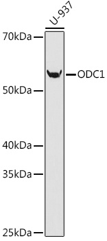
Western blot analysis of lysates from U-937 cells, using ODC1 Rabbit mAb (CAB3898) at 1:1000 dilution. Secondary antibody: HRP-conjugated Goat anti-Rabbit IgG (H+L) (CABS014) at 1:10000 dilution. Lysates/proteins: 25μg per lane. Blocking buffer: 3% nonfat dry milk in TBST. Detection: ECL Basic Kit (AbGn00020). Exposure time: 90s.


![Anti-ODC1 [R05-9Q7] Monoclonal Antibody (AGMB02649)](https://cdn11.bigcommerce.com/s-h68l9z2lnx/images/stencil/590x590/products/273938/676361/anti-odc1-r05-9q7-monoclonal-antibody-agmb02649__41137.1773029187.jpg?c=2 "Anti-ODC1 [R05-9Q7] Monoclonal Antibody (AGMB02649)")


