The SATB2 Monoclonal Antibody (CAB19837) is a high-quality antibody developed for reliable detection and analysis of target proteins. This antibody, produced using hybridoma technology, exhibits high specificity and sensitivity for SATB2 in human samples, making it suitable for various applications including immunohistochemistry and flow cytometry.SATB2 is known to play a key role in cell differentiation and development, particularly in the skeletal and nervous systems. Aberrant expression of SATB2 has been linked to various diseases, making it a potential therapeutic target for conditions like osteoporosis and neurological disorders.
This antibody is validated for use in WB, IHC-P, IP, ELISA applications and has demonstrated reactivity against Human, Mouse, Rat samples.
Product Name:
SATB2 Monoclonal Antibody
SKU:
CAB19837
Size:
20μL, 100μL
Reactivity:
Human, Mouse, Rat
Clone Number:
ARC2363
Conjugate:
Unconjugated
Immunogen:
Synthetic peptide. This information is considered to be commercially sensitive.
0.5μg-4μg antibody for 200μg-400μg extracts of whole cells
ELISA
Recommended starting concentration is 1 μg/mL. Please optimize the concentration based on your specific assay requirements.
Synonyms:
GLSS, DEL2Q32Q33, C2DELq32q33, SATB2
Positive Sample:
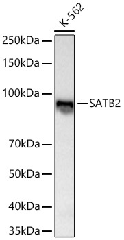
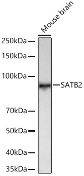
K-562, Mouse brain
Cellular Localization:
Nucleus.
Calculated MW:
83kDa
Observed MW:
100kDa
This gene encodes a DNA binding protein that specifically binds nuclear matrix attachment regions. The encoded protein is involved in transcription regulation and chromatin remodeling. Defects in this gene are associated with isolated cleft palate and cognitive disability. Alternate splicing results in multiple transcript variants that encode the same protein.
Purification Method
Affinity purification
Gene ID
23314
Buffer Information
Store at -20℃. Avoid freeze / thaw cycles. Buffer: PBS containing 50% glycerol and 0.05% BSA, preserved with proclin300 or sodium azide, pH 7.3.
Western blot analysis of lysates from K-562 cells using SATB2 Rabbit mAb (CAB19837) at 1:1000 dilution incubated overnight at 4℃. Secondary antibody: HRP-conjugated Goat anti-Rabbit IgG (H+L) (CABS014) at 1:10000 dilution. Lysates/proteins: 25 μg per lane. Blocking buffer: 3% nonfat dry milk in TBST. Detection: ECL Basic Kit (AbGn00020). Exposure time: 60s.
Western blot analysis of lysates from Mouse brain using SATB2 Rabbit mAb (CAB19837) at 1:1000 dilution incubated overnight at 4℃. Secondary antibody: HRP-conjugated Goat anti-Rabbit IgG (H+L) (CABS014) at 1:10000 dilution. Lysates/proteins: 25 μg per lane. Blocking buffer: 3% nonfat dry milk in TBST. Detection: ECL Basic Kit (AbGn00020). Exposure time: 90s.
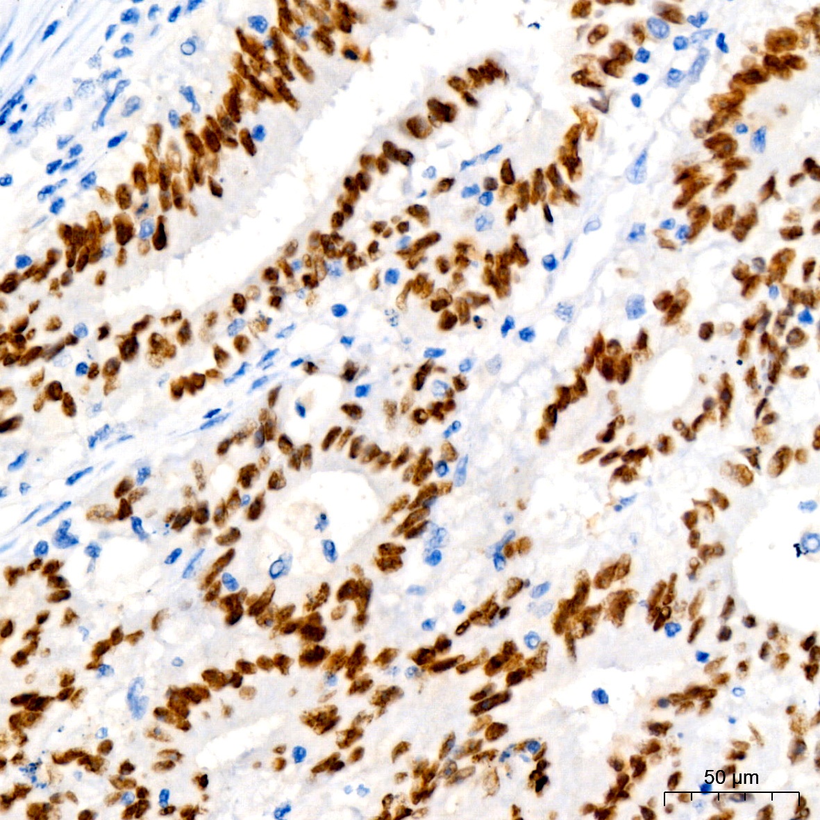
Immunohistochemistry analysis of paraffin-embedded Human colon carcinoma using SATB2 Rabbit mAb (CAB19837) at dilution of 1:50 (40x lens). High pressure antigen retrieval performed with 0.01M Tris/EDTA Buffer (pH 9.0) prior to IHC staining.
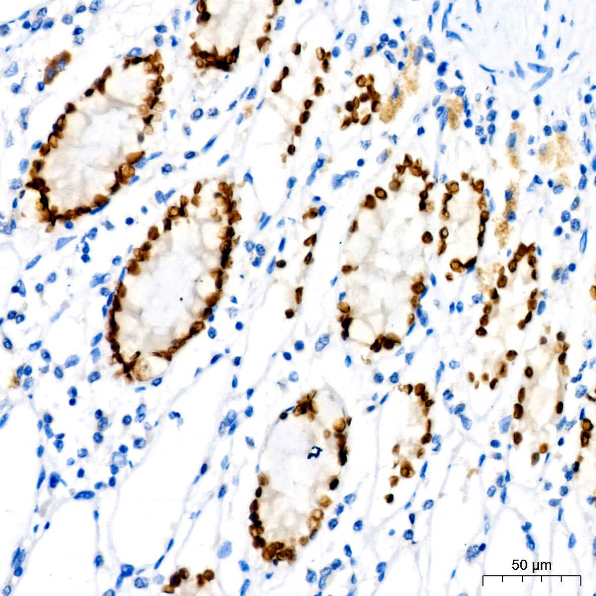
Immunohistochemistry analysis of paraffin-embedded Human colon using SATB2 Rabbit mAb (CAB19837) at dilution of 1:50 (40x lens). High pressure antigen retrieval performed with 0.01M Tris/EDTA Buffer (pH 9.0) prior to IHC staining.


![Anti-SATB2 [R06-6D8] Monoclonal Antibody (AGMB00083)](https://cdn11.bigcommerce.com/s-h68l9z2lnx/images/stencil/590x590/products/271372/692707/anti-satb2-r06-6d8-monoclonal-antibody-agmb00083__15938.1774507035.jpg?c=2 "Anti-SATB2 [R06-6D8] Monoclonal Antibody (AGMB00083)")
![Anti-SATB2 [R05-4K-3] Monoclonal Antibody (AGMB03734)](https://cdn11.bigcommerce.com/s-h68l9z2lnx/images/stencil/590x590/products/275023/676894/anti-satb2-r05-4k-3-monoclonal-antibody-agmb03734__13608.1773030869.jpg?c=2 "Anti-SATB2 [R05-4K-3] Monoclonal Antibody (AGMB03734)")


