The ARL3 Antibody (CAB16348) is a high-quality antibody developed for reliable detection and analysis of target proteins. Produced in rabbits, this antibody is highly specific to human ARL3 and has been validated for use in Western blotting applications. By binding to the ARL3 protein, researchers can accurately detect and analyze its expression in various cell types, making it an ideal reagent for studies in cell biology and developmental biology.
This antibody is validated for use in WB, IF/ICC, ELISA applications and has demonstrated reactivity against Human, Mouse, Rat samples.
Product Name:
ARL3 Antibody
SKU:
CAB16348
Size:
20μL, 100μL
Reactivity:
Human, Mouse, Rat
Conjugate:
Unconjugated
Immunogen:
Recombinant protein (or fragment).This information is considered to be commercially sensitive.
Enables GDP binding activity; GTP binding activity; and microtubule binding activity. Involved in several processes, including cilium assembly; protein localization to cilium; and small GTPase mediated signal transduction. Acts upstream of or within post-Golgi vesicle-mediated transport. Located in several cellular components, including microtubule cytoskeleton; midbody; and photoreceptor connecting cilium. Implicated in Joubert syndrome and retinitis pigmentosa 83.
Purification Method
Affinity purification
Gene ID
403
RRID
AB_2768440
Buffer Information
Store at -20℃. Avoid freeze / thaw cycles. Buffer: PBS with 0.01% thimerosal,50% glycerol,pH7.3.
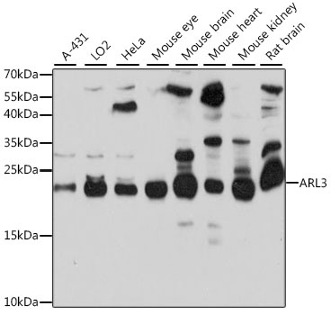
Western blot analysis of various lysates using ARL3 Rabbit pAb (CAB16348) at 1:1000 dilution. Secondary antibody: HRP-conjugated Goat anti-Rabbit IgG (H+L) (CABS014) at 1:10000 dilution. Lysates/proteins: 25μg per lane. Blocking buffer: 3% nonfat dry milk in TBST. Detection: ECL Basic Kit (AbGn00020). Exposure time: 30s.
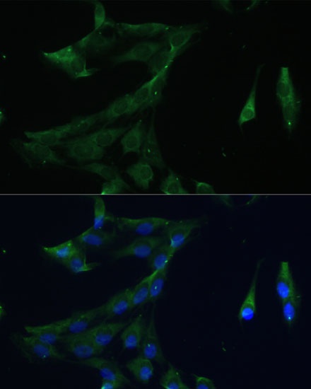
Immunofluorescence analysis of C6 cells using ARL3 Rabbit pAb (CAB16348) at dilution of 1:100 (40x lens). Secondary antibody: Cy3-conjugated Goat anti-Rabbit IgG (H+L) (CABS007) at 1:500 dilution. Blue: DAPI for nuclear staining.