The ATF5 Antibody (CAB18155) is a high-quality antibody developed for reliable detection and analysis of target proteins. This rabbit polyclonal antibody is highly specific for ATF5 in human samples and is validated for use in Western blot applications. By binding to the ATF5 protein, this antibody enables researchers to detect and analyze ATF5 expression in a variety of cell types, making it ideal for studies in cell biology, cancer research, and neurobiology.
This antibody is validated for use in WB, IHC-P, IF/ICC, ELISA applications and has demonstrated reactivity against Human, Mouse, Rat samples.
Product Name:
ATF5 Antibody
SKU:
CAB18155
Size:
20μL, 100μL
Reactivity:
Human, Mouse, Rat
Immunogen:
Recombinant protein (or fragment).This information is considered to be commercially sensitive.
Recommended starting concentration is 1 μg/mL. Please optimize the concentration based on your specific assay requirements.
Synonyms:
ATFX, HMFN0395, ATF5
Positive Sample:
rat lung
Cellular Localization:
Centrosome, Cytosol, Nucleoplasm, Nucleus.
Calculated MW:
31kDa
Observed MW:
31kDa
Enables several functions, including DNA-binding transcription activator activity, RNA polymerase II-specific; RNA polymerase II transcription regulatory region sequence-specific DNA binding activity; and tubulin binding activity. Involved in several processes, including fat cell differentiation; regulation of cell cycle process; and regulation of transcription, DNA-templated. Located in centrosome; cytosol; and nucleoplasm.
Purification Method
Affinity purification
Gene ID
22809
RRID
AB_2861945
Buffer Information
Store at -20℃. Avoid freeze / thaw cycles. Buffer: Buffer: PBS containing 50% glycerol, preserved with proclin300 or sodium azide, pH 7.3.
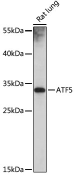
Western blot analysis of lysates from rat lung, using ATF5 Rabbit pAb (CAB18155) at 1:1000 dilution. Secondary antibody: HRP-conjugated Goat anti-Rabbit IgG (H+L) (CABS014) at 1:10000 dilution. Lysates/proteins: 25μg per lane. Blocking buffer: 3% nonfat dry milk in TBST. Detection: ECL Basic Kit (AbGn00020). Exposure time: 10s.
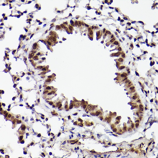
Immunohistochemistry analysis of paraffin-embedded Mouse lung using ATF5 Rabbit pAb (CAB18155) at dilution of 1:20 (40x lens). High pressure antigen retrieval performed with 0.01M Citrate buffer (pH 6.0) prior to IHC staining.
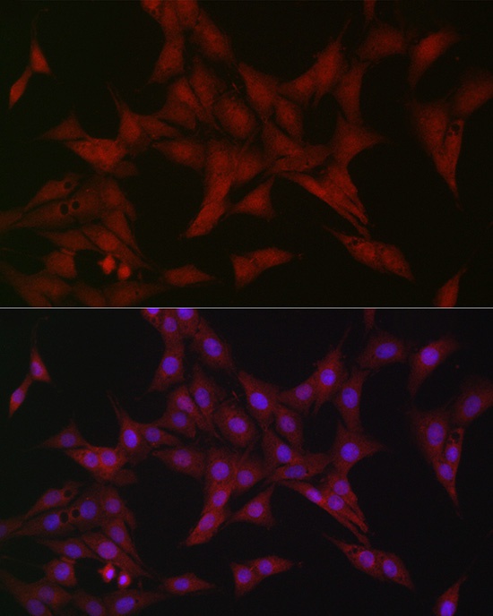
Immunofluorescence analysis of PC-12 cells using ATF5 Rabbit pAb (CAB18155) at dilution of 1:20 (40x lens). Secondary antibody: Cy3-conjugated Goat anti-Rabbit IgG (H+L) (CABS007) at 1:500 dilution. Blue: DAPI for nuclear staining.
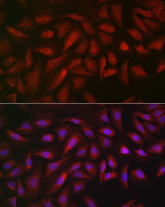
Immunofluorescence analysis of U2OS cells using ATF5 Rabbit pAb (CAB18155) at dilution of 1:20 (40x lens). Secondary antibody: Cy3-conjugated Goat anti-Rabbit IgG (H+L) (CABS007) at 1:500 dilution. Blue: DAPI for nuclear staining.