The BRPF1 Antibody (CAB17012) is a high-quality antibody developed for reliable detection and analysis of target proteins. This antibody, generated in rabbits, demonstrates high specificity and sensitivity when detecting BRPF1 in human samples, making it ideal for use in Western blot applications.BRPF1 is essential for chromatin remodeling and transcriptional regulation, playing a vital role in cellular processes such as differentiation and development.
This antibody is validated for use in WB, IF/ICC, ELISA applications and has demonstrated reactivity against Human, Mouse, Rat samples.
Product Name:
BRPF1 Antibody
SKU:
CAB17012
Size:
20μL, 100μL
Reactivity:
Human, Mouse, Rat
Immunogen:
Recombinant protein (or fragment).This information is considered to be commercially sensitive.
This gene encodes a bromodomain, PHD finger and chromo/Tudor-related Pro-Trp-Trp-Pro (PWWP) domain containing protein. The encoded protein is a component of the MOZ/MORF histone acetyltransferase complexes which function as a transcriptional regulators. This protein binds to the catalytic MYST domains of the MOZ and MORF proteins and may play a role in stimulating acetyltransferase and transcriptional activity of the complex. Alternative splicing results in multiple transcript variants.
Purification Method
Affinity purification
Gene ID
7862
RRID
AB_2768624
Buffer Information
Store at -20℃. Avoid freeze / thaw cycles. Buffer: PBS with 0.01% thimerosal,50% glycerol,pH7.3.
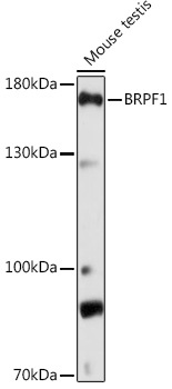
Western blot analysis of lysates from Mouse testis, using BRPF1 Rabbit pAb (CAB17012) at 1:1000 dilution. Secondary antibody: HRP-conjugated Goat anti-Rabbit IgG (H+L) (CABS014) at 1:10000 dilution. Lysates/proteins: 25μg per lane. Blocking buffer: 3% nonfat dry milk in TBST. Detection: ECL Basic Kit (AbGn00020). Exposure time: 90s.
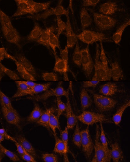
Immunofluorescence analysis of C6 cells using BRPF1 Rabbit pAb (CAB17012) at dilution of 1:100. Secondary antibody: Cy3-conjugated Goat anti-Rabbit IgG (H+L) (CABS007) at 1:500 dilution. Blue: DAPI for nuclear staining.
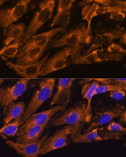
Immunofluorescence analysis of U-2 OS cells using BRPF1 Rabbit pAb (CAB17012) at dilution of 1:100. Secondary antibody: Cy3-conjugated Goat anti-Rabbit IgG (H+L) (CABS007) at 1:500 dilution. Blue: DAPI for nuclear staining.