The CA5A Antibody (CAB14709) is a high-quality antibody developed for reliable detection and analysis of target proteins. This antibody, produced in rabbits, shows high reactivity with human samples and has been validated for use in Western blot applications. By binding specifically to the CA5A protein, this antibody enables the detection and analysis of CA5A in various cell types, making it ideal for investigations in physiology and disease research.CA5A, also known as carbonic anhydrase VA, plays a crucial role in maintaining acid-base balance and pH regulation in tissues.
This antibody is validated for use in WB, IF/ICC, ELISA applications and has demonstrated reactivity against Human, Mouse, Rat samples.
Product Name:
CA5A Antibody
SKU:
CAB14709
Size:
20μL, 100μL
Reactivity:
Human, Mouse, Rat
Conjugate:
Unconjugated
Immunogen:
Recombinant protein (or fragment).This information is considered to be commercially sensitive.
Recommended starting concentration is 1 μg/mL. Please optimize the concentration based on your specific assay requirements.
Synonyms:
CA5, CAV, CAVA, CA5AD, GS1-21A4.1, CA5A
Positive Sample:
Mouse liver
Cellular Localization:
Mitochondrion.
Calculated MW:
35kDa
Observed MW:
36kDa
Carbonic anhydrases (CAs) are a large family of zinc metalloenzymes that catalyze the reversible hydration of carbon dioxide. They participate in a variety of biological processes, including respiration, calcification, acid-base balance, bone resorption, and the formation of aqueous humor, cerebrospinal fluid, saliva, and gastric acid. They show extensive diversity in tissue distribution and in their subcellular localization. CA VA is localized in the mitochondria and expressed primarily in the liver. It may play an important role in ureagenesis and gluconeogenesis. CA5A gene maps to chromosome 16q24.3 and an unprocessed pseudogene has been assigned to 16p12-p11.2.
Purification Method
Affinity purification
Gene ID
763
RRID
AB_2761584
Buffer Information
Store at -20℃. Avoid freeze / thaw cycles. Buffer: PBS containing 50% glycerol, preserved with proclin300 or sodium azide, pH 7.3.
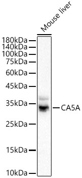
Western blot analysis of lysates from Mouse liver using CA5A Rabbit pAb (CAB14709) at 1:1000 dilution. Secondary antibody: HRP-conjugated Goat anti-Rabbit IgG (H+L) (CABS014) at 1:10000 dilution. Lysates/proteins: 25 μg per lane. Blocking buffer: 3% nonfat dry milk in TBST. Detection: ECL Basic Kit (AbGn00020). Exposure time:5s.
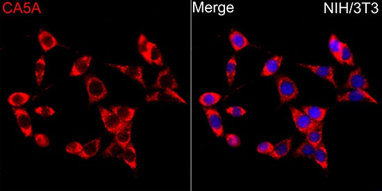
Immunofluorescence analysis of NIH/3T3 cells using CA5A Rabbit pAb (CAB14709) at dilution of 1:100 (40x lens). Secondary antibody: Cy3-conjugated Goat anti-Rabbit IgG (H+L) (CABS007) at 1:500 dilution. Blue: DAPI for nuclear staining.