The CBL Antibody (CAB0732) is a high-quality antibody developed for reliable detection and analysis of target proteins. This antibody, generated in rabbits, demonstrates high reactivity with human samples and has been validated for use in Western blot applications. By binding to the CBL protein, this antibody enables precise detection and analysis in a variety of cell types, making it well-suited for studies in cancer research and cell signaling.CBL is known for its role in regulating cell growth, division, and differentiation, making it a key player in various cellular processes.
This antibody is validated for use in WB, IHC-P, IF/ICC, IP, ELISA applications and has demonstrated reactivity against Human, Mouse, Rat samples.
Product Name:
CBL Antibody
SKU:
CAB0732
Size:
20μL, 100μL
Reactivity:
Human, Mouse, Rat
Conjugate:
Unconjugated
Immunogen:
Recombinant protein (or fragment).This information is considered to be commercially sensitive.
0.5μg-4μg antibody for 400μg-600μg extracts of whole cells
ELISA
Recommended starting concentration is 1 μg/mL. Please optimize the concentration based on your specific assay requirements.
Synonyms:
CBL2, NSLL, C-CBL, RNF55, FRA11B, CBL
Positive Sample:
293T, Mouse thymus
Cellular Localization:
Cell Membrane, Cytoplasm.
Calculated MW:
100kDa
Observed MW:
120kDa/110kDa
This gene is a proto-oncogene that encodes a RING finger E3 ubiquitin ligase. The encoded protein is one of the enzymes required for targeting substrates for degradation by the proteasome. This protein mediates the transfer of ubiquitin from ubiquitin conjugating enzymes (E2) to specific substrates. This protein also contains an N-terminal phosphotyrosine binding domain that allows it to interact with numerous tyrosine-phosphorylated substrates and target them for proteasome degradation. As such it functions as a negative regulator of many signal transduction pathways. This gene has been found to be mutated or translocated in many cancers including acute myeloid leukaemia, and expansion of CGG repeats in the 5' UTR has been associated with Jacobsen syndrome. Mutations in this gene are also the cause of Noonan syndrome-like disorder.
Purification Method
Affinity purification
Gene ID
867
RRID
AB_2757369
Buffer Information
Store at -20℃. Avoid freeze / thaw cycles. Buffer: PBS containing 50% glycerol, preserved with proclin300 or sodium azide, pH 7.3.
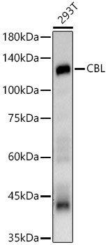
Western blot analysis of lysates from 293T cells, using CBL Rabbit pAb (CAB0732) at 1:3000 dilution. Secondary antibody: HRP-conjugated Goat anti-Rabbit IgG (H+L) (CABS014) at 1:10000 dilution. Lysates/proteins: 25μg per lane. Blocking buffer: 3% nonfat dry milk in TBST. Detection: ECL Basic Kit (AbGn00020). Exposure time: 30s.
Western blot analysis of lysates from Mouse thymus, using CBL Rabbit pAb (CAB0732) at 1:3000 dilution. Secondary antibody: HRP-conjugated Goat anti-Rabbit IgG (H+L) (CABS014) at 1:10000 dilution. Lysates/proteins: 25μg per lane. Blocking buffer: 3% nonfat dry milk in TBST. Detection: ECL Basic Kit (AbGn00020). Exposure time: 1s.
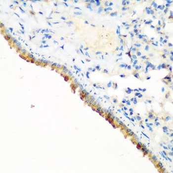
Immunohistochemistry analysis of paraffin-embedded Mouse lung using CBL Rabbit pAb (CAB0732) at dilution of 1:200 (40x lens). Microwave antigen retrieval performed with 0.01M PBS Buffer (pH 7.2) prior to IHC staining.
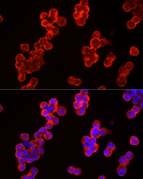
Immunofluorescence analysis of K-562 cells using CBL Rabbit pAb (CAB0732) at dilution of 1:300 (40x lens). Secondary antibody: Cy3-conjugated Goat anti-Rabbit IgG (H+L) (CABS007) at 1:500 dilution. Blue: DAPI for nuclear staining.
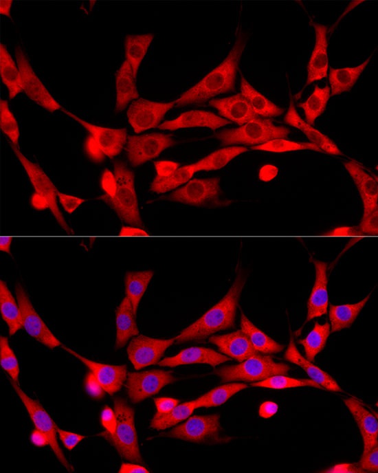
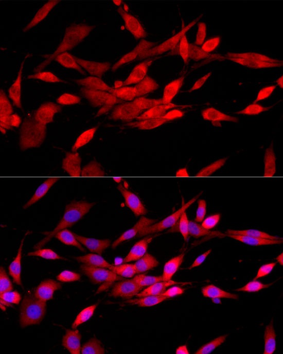
Immunofluorescence analysis of PC-12 cells using CBL Rabbit pAb (CAB0732) at dilution of 1:300 (40x lens). Secondary antibody: Cy3-conjugated Goat anti-Rabbit IgG (H+L) (CABS007) at 1:500 dilution. Blue: DAPI for nuclear staining.
Immunofluorescence analysis of PC-12 cells using CBL Rabbit pAb (CAB0732) at dilution of 1:300 (40x lens). Secondary antibody: Cy3-conjugated Goat anti-Rabbit IgG (H+L) (CABS007) at 1:500 dilution. Blue: DAPI for nuclear staining.
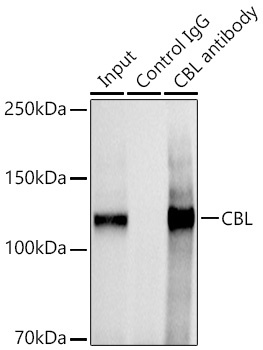
Immunoprecipitation analysis of 600 μg extracts of Mouse thymus cells using 3 μg CBL antibody (CAB0732). Western blot was performed from the immunoprecipitate using CBL antibody at a dilution of 1:1000.